. 2022 Jun 28;13(3):e0034222.
doi: 10.1128/mbio.00342-22. Epub 2022 May 16.

Blocking Polyphosphate Mobilization Inhibits Pho4 Activation and Virulence in the Pathogen Candida albicans

Affiliations

Blocking Polyphosphate Mobilization Inhibits Pho4 Activation and Virulence in the Pathogen Candida albicans

Yasmin Ahmed et al. mBio. .

Abstract

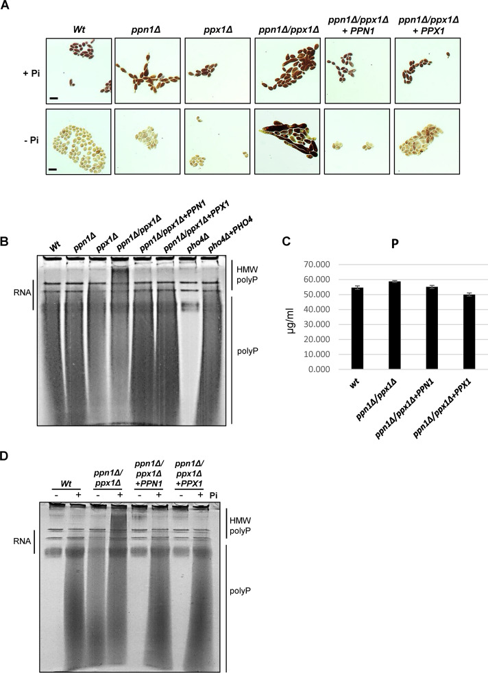
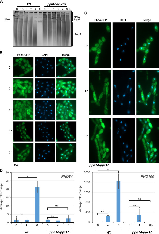
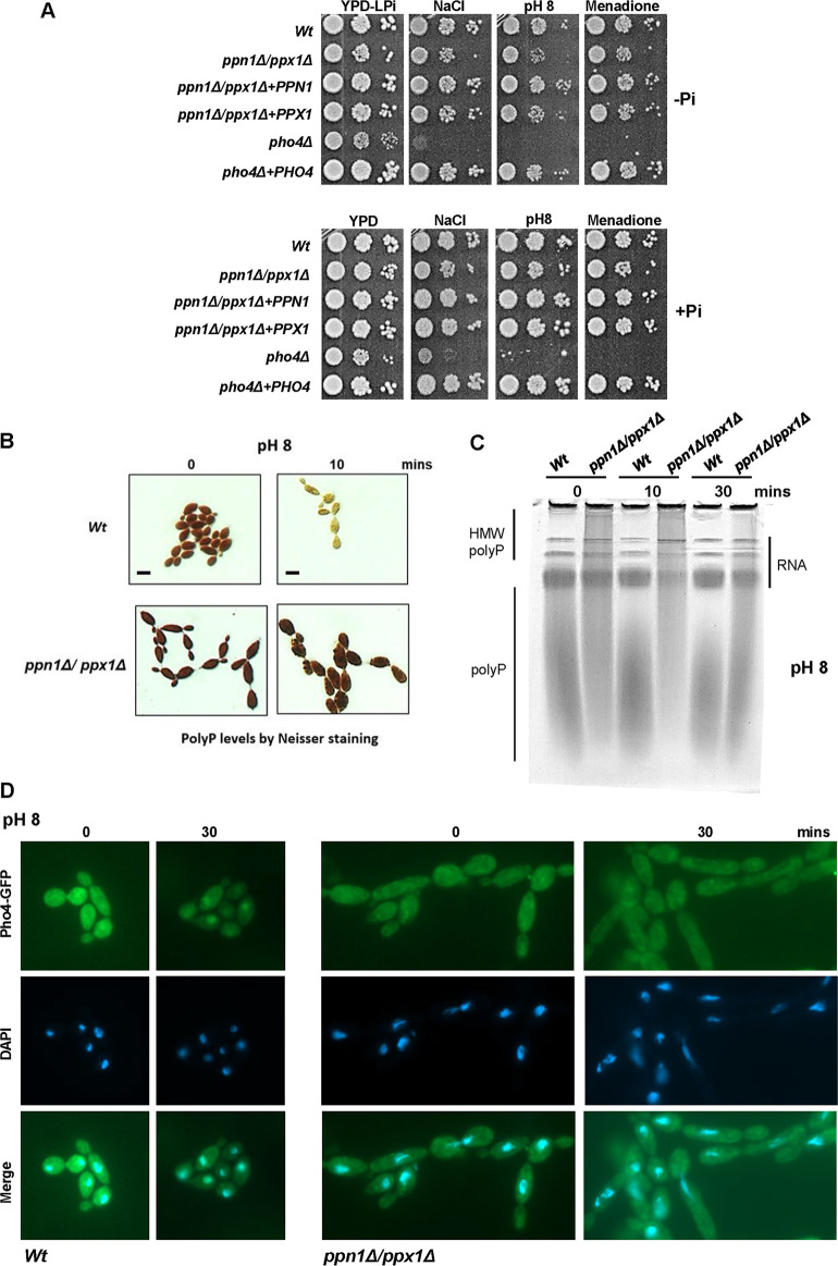
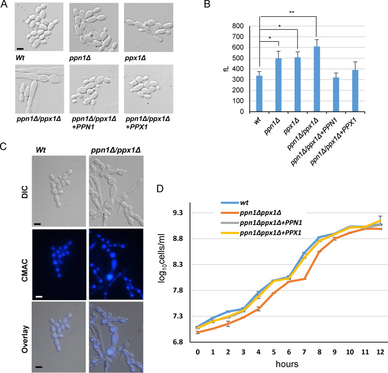
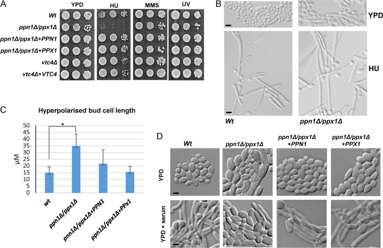
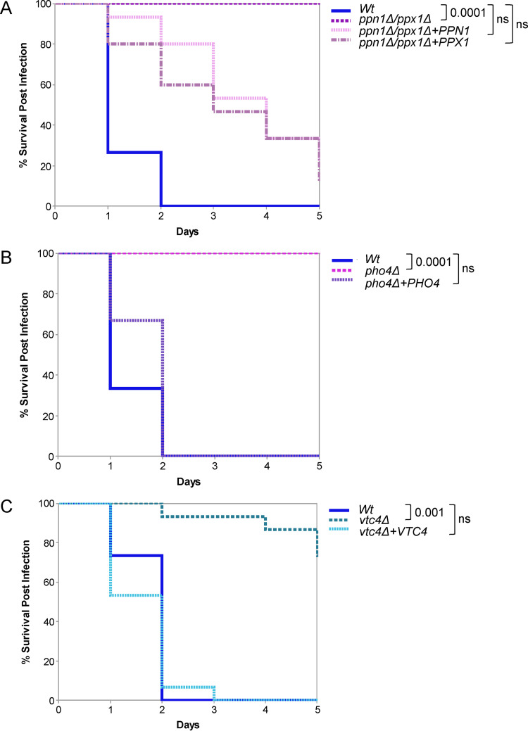
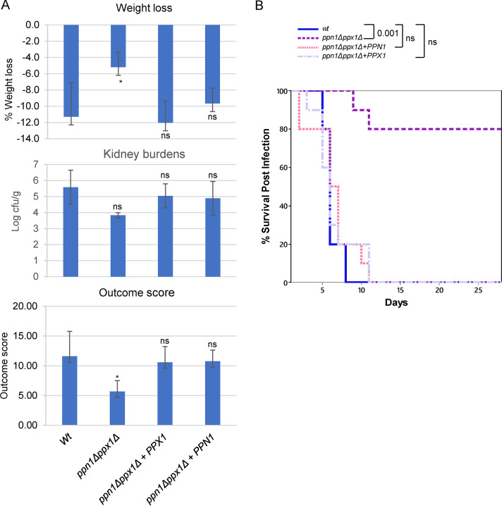
The ability of pathogenic fungi to obtain essential nutrients from the host is vital for virulence. In Candida albicans, acquisition of the macronutrient phosphate is regulated by the Pho4 transcription factor and is important for both virulence and resistance to host-encountered stresses. All cells store phosphate in the form of polyphosphate (polyP), a ubiquitous polymer comprising tens to hundreds of phosphate residues. Release of phosphate from polyP is one of the first responses evoked in response to phosphate starvation, and here, we sought to explore the importance of polyP mobilization in the pathobiology of C. albicans. We found that two polyphosphatases, Ppn1 and Ppx1, function redundantly to release phosphate from polyP in C. albicans. Strikingly, we reveal that blocking polyP mobilization prevents the activation of the Pho4 transcription factor: following Pi starvation, Pho4 fails to accumulate in the nucleus and induce Pi acquisition genes in ppn1Δ ppx1Δ cells. Consequently, ppn1Δ ppx1Δ cells display impaired resistance to the same range of stresses that require Pho4 for survival. In addition, cells lacking both polyphosphatases are exquisitely sensitive to DNA replication stress, indicating that polyP mobilization is needed to support the phosphate-demanding process of DNA replication. Blocking polyP mobilization also results in significant morphological defects, as ppn1Δ ppx1Δ cells form large pseudohypha-like cells that are resistant to serum-induced hypha formation. Thus, polyP mobilization impacts key processes important for the pathobiology of C. albicans, and consistent with this, we found that blocking this process attenuates the virulence of this important human fungal pathogen. IMPORTANCE Acquisition of the essential macronutrient phosphate is important for the virulence of Candida albicans, a major human fungal pathogen. All cells store phosphate as polyphosphate (polyP), which is rapidly mobilized when phosphate is limiting. Here, we identified the major phosphatases involved in releasing phosphate from polyP in C. albicans. By blocking this process, we found that polyP mobilization impacts many process that contribute to C. albicans pathogenesis. Notably, we found that blocking polyP mobilization inhibits activation of the Pho4 transcription factor, the master regulator of phosphate acquisition. In addition, cell cycle progression, stress resistance, morphogenetic switching, and virulence are all impaired in cells that cannot mobilize polyP. This study therefore provides new insight into the importance of polyP mobilization in promoting the virulence of C. albicans. As phosphate homeostasis strategies differ between fungal pathogen and host, this offers promise for the future development of antifungals.

Keywords: Candida albicans; morphogenesis; phosphate metabolism; stress response; virulence.


Conflict of interest statement

The authors declare no conflict of interest.

Figures

FIG 1
FIG 1
Ppn1 and Ppx1 function redundantly to mobilize polyP stores. (A) Neisser staining of the indicated strains following growth in YPD+Pi or YPD-LPi for 16 h. Bars represent 10 μm, and the scale is the same across each row of images. (B) Toluidine blue staining of RNA/polyP extracts after electrophoresis on urea-polyacrylamide gels from cells grown in YPD. The high-molecular-weight (HMW) polyP in ppn1Δ ppx1Δ cells is indicated. (C) Impact of PPX1 and PPN1 loss on intracellular phosphate levels. Whole-cell nitric acid digests of WT, ppn1Δ ppx1Δ, ppn1Δ ppx1Δ+PPN1, and ppn1Δ ppx1Δ+PPX1 cells grown in YPD were analyzed by ICP-MS. Phosphate levels shown are means and SD for three independent cultures. (D) Toluidine blue staining of RNA/polyP extracts after electrophoresis on urea-polyacrylamide gels from cells grown in YPD+Pi or YPD-LPi for 16 h.
FIG 2
FIG 2
Pho4 activation in ppn1Δ ppx1Δ cells. (A) Toluidine blue staining of RNA/polyP extracts after electrophoresis on urea-polyacrylamide gels from WT and ppn1Δ ppx1Δ cells expressing Pho4-GFP grown in YPD until mid-log phase (t = 0) and then moved to YPD-LPi medium for the indicated times. (B and C) Cells from the cultures described above were processed to examine Pho4-GFP localization using fluorescence microscopy. DAPI staining illustrates nuclear positioning. (D) RT-qPCR analysis showing fold induction of the Pho4 target genes PHO84 and PHO100 after growth for 4 and 6 h in YPD-LPi. Transcript levels were measured relative to the internal ACT1 mRNA control and normalized to the level of transcript in WT cells with Pi. Means and standard deviations for three biological replicates are shown. ns, not significant; *, P < 0.05; **, P < 0.01.
FIG 3
FIG 3
PolyP mobilization and stress responses. (A) PolyP mobilization promotes stress resistance under Pi-limiting conditions. Exponentially growing strains were spotted in serial dilutions onto YPD-LPi plates containing 1 M NaCl and 300 μM menadione or at pH 8 (top, −Pi) or in YPD plates containing the same stresses (bottom, +Pi). Plates were incubated for 24 h at 30°C. (B) PolyP mobilization in response to alkaline stress is dependent on Ppn1 and Ppx1. Neisser staining of the indicated strains grown in YPD or after 10 min growth in YPD (pH 8). Bar, 10 μm. (C) Toluidine blue staining of RNA/polyP extracts from indicated strains after electrophoresis on urea-polyacrylamide gels, before and after 30 min growth in YPD medium (pH 8). (D) WT and ppn1Δ ppx1Δ cells expressing Pho4-GFP were grown in YPD, left untreated or transferred to YPD medium (pH 8) for 30 min, and processed to examine Pho4-GFP localization using fluorescence microscopy. DAPI staining illustrates nuclear positioning.
FIG 4
FIG 4
Morphological and growth characteristics of ppn1Δ ppx1Δ cells. (A) Cells lacking Ppn1 and Ppx1 have altered morphologies. DIC images of exponentially growing cells. (B) Cells lacking Ppn1 and Ppx1 are larger than wild-type cells. Cell volume analysis showing the cell volumes (means and SD). The data were analyzed statistically using Student’s two-sample t test. ns, not significant; *, P < 0.05; **, P < 0.01. (C) Some ppn1Δ ppx1Δ cells exhibit large vacuoles. Vacuolar morphology was captured by CMAC staining. (D) Cells lacking PPN1 and PPX1 have a slight slow-growth phenotype. Analysis of growth of the indicated strains in YPD. Cells numbers rather than OD were recorded due to the morphological defects seen in ppn1Δ ppx1Δ cells. Bars (A and C) represent 10 μm, and scale is the same across each row of images.
FIG 5
FIG 5
Ppn1 and Ppx1 are required for replication stress resistance and the formation of hyphae. (A) Exponentially growing strains were spotted in serial dilutions onto YPD plates that contained HU (40 mM) or MMS (0.02%) or that were exposed to UV (75 J/m2). Plates were incubated for 24 h at 30°C. (B) DIC images of cells grown in YPD and following treatment with 40 mM HU for 4 h. Bars represent 10 μm, and scale is the same across each row of images. (C) Quantification of hyperpolarized bud length was carried out using Zeiss imaging software on 200 cells for each strain. Data are means and SD. Statistical analysis was performed using Student’s two-sample t test. *, P < 0.05. (D) Stationary-phase cells were diluted 1:10 in YPD medium containing 10% fetal bovine serum and incubated at 37°C for 3 h (YPD + serum). Bars represent 10 μm, and scale is the same across each row of images.
FIG 6
FIG 6
Virulence analysis in the Galleria mellonella model of infection. (A to C) Comparison of virulence of the indicated strains in the Galleria model of systemic infection (15 larvae per fungal strain). The data were analyzed statistically using the log-rank (Mantel-Cox) test. ns, not significant.
FIG 7
FIG 7
Virulence analysis in murine models of infection. (A) Three-day infection model. Kidney burden, percentage weight loss, and outcome scores for mice (n = 6) infected with the indicated strains. Comparison of WT, ppn1Δ ppx1Δ+PPN1, and ppn1Δ ppx1Δ+PPX1 strain-infected mice with ppn1Δ ppx1Δ strain-infected mice by Kruskal-Wallis statistical analysis demonstrated a significant difference with ppn1Δ ppx1Δ strain-infected mice across all three parameters. ns, not significant; *, P < 0.05. (B) Twenty-eight-day survival model. Mice were injected with the same strains as for panel A, and survival was monitored daily. Survival curves were created using 10 mice per group except for the ppn1Δ ppx1Δ+PPX1 strain (n = 9). Comparing survival of each strain with WT cells, only ppn1Δ ppx1Δ cells were highly significantly different (Kruskal-Wallis nonparametric test).
FIG 8
FIG 8
Activation of Pho4 is inhibited when polyP mobilization is hindered. Growth of wild-type C. albicans cells in Pi-limiting medium stimulates polyP mobilization, the nuclear accumulation of Pho4, and the induction of Pho4-dependent genes with roles in Pi acquisition. However, in cells lacking the Ppn1 and Ppx1 polyphosphatases, polyP mobilization is dramatically impaired following growth in Pi-limiting medium. Furthermore, Pho4 fails to accumulate in the nucleus and activate Pi acquisition genes. This suggests that an inability to mobilize polyP in C. albicans prevents the activation of Pho4. In S. cerevisiae, Pho4 is activated following Pi limitation via Pho81 inhibition of the Pho80-Pho85 cyclin-CDK complex, which negatively regulates Pho4. It is unknown if C. albicans Pho4 is regulated in the same way (as indicated by the dashed lines), but it is possible that polyP presence interferes with mechanism by which the cell senses Pi-limiting environments. The figure was created with BioRender.com.

Similar articles

Cited by

References

    1. Brown GD, Denning DW, Gow NA, Levitz SM, Netea MG, White TC. 2012. Hidden killers: human fungal infections. Sci Transl Med 4:165rv13. doi:10.1126/scitranslmed.3004404. - DOI - PubMed
    1. Ene IV, Brunke S, Brown AJ, Hube B. 2014. Metabolism in fungal pathogenesis. Cold Spring Harb Perspect Med 4:a019695. doi:10.1101/cshperspect.a019695. - DOI - PMC - PubMed
    1. Citiulo F, Jacobsen ID, Miramón P, Schild L, Brunke S, Zipfel P, Brock M, Hube B, Wilson D. 2012. Candida albicans scavenges host zinc via Pra1 during endothelial invasion. PLoS Pathog 8:e1002777. doi:10.1371/journal.ppat.1002777. - DOI - PMC - PubMed
    1. Ikeh M, Ahmed Y, Quinn J. 2017. Phosphate acquisition and virulence in human fungal pathogens. Microorganisms 5:48. doi:10.3390/microorganisms5030048. - DOI - PMC - PubMed
    1. Ikeh MAC, Kastora SL, Day AM, Herrero-de-Dios CM, Tarrant E, Waldron KJ, Banks AP, Bain JM, Lydall D, Veal EA, MacCallum DM, Erwig LP, Brown AJP, Quinn J. 2016. Pho4 mediates phosphate acquisition in Candida albicans and is vital for stress resistance and metal homeostasis. Mol Biol Cell 27:2784–2801. doi:10.1091/mbc.e16-05-0266. - DOI - PMC - PubMed

Publication types

MeSH terms