. 2022 Jun 8;96(11):e0063422.
doi: 10.1128/jvi.00634-22. Epub 2022 May 16.

Endogenous Retroviruses Augment Amphibian (Xenopus laevis) Tadpole Antiviral Protection

Affiliations

Endogenous Retroviruses Augment Amphibian (Xenopus laevis) Tadpole Antiviral Protection

Namarta Kalia et al. J Virol. .

Abstract

The global amphibian declines are compounded by infections with members of the Ranavirus genus such as Frog Virus 3 (FV3). Premetamorphic anuran amphibians are believed to be significantly more susceptible to FV3 while this pathogen targets the kidneys of both pre- and postmetamorphic animals. Paradoxically, FV3-challenged Xenopus laevis tadpoles exhibit lower kidney viral loads than adult frogs. Presently, we demonstrate that X. laevis tadpoles are intrinsically more resistant to FV3 kidney infections than cohort-matched metamorphic and postmetamorphic froglets and that this resistance appears to be epigenetically conferred by endogenous retroviruses (ERVs). Using a X. laevis kidney-derived cell line, we show that enhancing ERV gene expression activates cellular double-stranded RNA-sensing pathways, resulting in elevated mRNA levels of antiviral interferon (IFN) cytokines and thus greater anti-FV3 protection. Finally, our results indicate that large esterase-positive myeloid-lineage cells, rather than renal cells, are responsible for the elevated ERV/IFN axis seen in the tadpole kidneys. This conclusion is supported by our observation that CRISPR-Cas9 ablation of colony-stimulating factor-3 results in abolished homing of these myeloid cells to tadpole kidneys, concurrent with significantly abolished tadpole kidney expression of both ERVs and IFNs. We believe that the manuscript marks an important step forward in understanding the mechanisms controlling amphibian antiviral defenses and thus susceptibility and resistance to pathogens like FV3. IMPORTANCE Global amphibian biodiversity is being challenged by pathogens like the Frog Virus 3 (FV3) ranavirus, underlining the need to gain a greater understanding of amphibian antiviral defenses. While it was previously believed that anuran (frog/toad) amphibian tadpoles are more susceptible to FV3, we demonstrated that tadpoles are in fact more resistant to this virus than metamorphic and postmetamorphic froglets. We showed that this resistance is conferred by large myeloid cells within the tadpole kidneys (central FV3 target), which possess an elevated expression of endogenous retroviruses (ERVs). In turn, these ERVs activate cellular double-stranded RNA-sensing pathways, resulting in a greater expression of antiviral interferon cytokines, thereby offering the observed anti-FV3 protection.

Keywords: amphibian immunity; antiviral responses; endogenous retroviruses; interferon response.


Conflict of interest statement

The authors declare no conflict of interest.

Figures
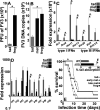
FIG 1
FIG 1
Tadpole kidney resistance to FV3 is marked by intrinsic antiviral protection. (A) Plaque assay analysis of FV3-infected tadpole and froglet kidneys (n = 6) at 6 h postinfection (hpi). (B) FV3 DNA viral loads in X. laevis tadpoles, metamorphs (NF 64), and froglets (n = 5/developmental stage). (C and D) IFN (C) and IRF (D) gene expression in healthy tadpole (NF 54) and froglet kidneys (n = 6/developmental stage). (E) Survival of tadpole (NF 54), early (NF 58) and late (NF 64) metamorphs, and froglets (n = 12/developmental stage) following FV3 infections (2.5 × 105 PFU of FV3/animal). (F) Percent tail absorption in metamorphosing tadpoles infected with WT- or Δ52L-FV3 for 4 days (2.5 × 105 PFU of FV3/animal; n = 9 per group). Results in A to D and F are means + SEM. Gene expression (C and D) was assessed relative to the gapdh endogenous control. Asterisks (*) above lines indicate statistical differences between treatment groups indicated by those lines.
FIG 2
FIG 2
The antiviral state within tadpole kidneys is conferred by elevated ERV expression. (A) Quantitative analysis of ERV gene expression in kidneys of healthy tadpoles (NF 54), metamorphic animals (NF 64), and froglets (n = 6/stage). (B and C) ERV (B) and IFN (C) gene expression in A6 cells treated with the Aza DNA methylation inhibitors (dissolved in APBS) or solvent control (APBS n = 6/treatment group). (D and E) Xen1 (D) and IFN (E) gene expression in A6 cells transfected with a Xen1 expression construct or an empty expression vector (vect; n = 6). (F) FV3 DNA loads in empty plasmid- and Xen1 construct-transfected A6 cells 24 h after FV3 infection (MOI of 0.5; n = 6). (G) Xen1, IFN7, and IFNL3 gene expression in A6 cells overexpressing Xen1, transfected with anti-MAVS morpholino (n = 6). Gene expression (A to E and G) was assessed relative to the gapdh endogenous control. Results are means + SEM. Asterisks (*) above lines indicate statistical differences between treatment groups indicated by those lines.
FIG 3
FIG 3
Esterase-positive myeloid cells are responsible for the high kidney ERV and IFN expression. Tadpole (NF 54) (A and B) and froglet (D) kidneys were stained and examined for the relative presence of esterase-positive (granulocyte/monocyte marker) cells. (C) Esterase-positive myeloid cells from tadpole kidneys. The results in A to D are representative of histological analyses of kidneys from 6 different tadpoles or froglets (n = 6/stage). (E) Tadpole kidney cell suspensions were subjected to chemotaxis assays against rCSF3 (100 to 10−8 ng/mL), using 5 × 105 cells per well of chemotaxis chambers (n = 4 to 8). Chemokinesis (chemo-kin) was assessed by examining migration with 104 ng/mL of rCSF3 in lower and upper chemotaxis chambers (n = 6). The role of kidney cell CSF3R in the rCSF3-mediated chemotaxis was examined by assessing migration in the absence or presence of 5 μg/mL of a soluble (extracellular portion of) rCSF3R (n = 6). (F) Comparison of Xen1 gene expression in tadpole rCSF3-recruited kidney cells (n = 6) relative to other X. laevis myeloid cells (n = 5). (G) Xen1 and IFN gene expression in total and rCSF3-chemoattracted tadpole kidney cells (n = 6). The results in E to G are means + SEM. Gene expression (F and G) was assessed relative to the gapdh endogenous control. Arrows in (A) and (D) indicate esterase-positive cells. Asterisks (*) above lines indicate statistical differences between treatment groups indicated by those lines. The diamond symbol (♦) indicates statistical difference from all other groups.
FIG 4
FIG 4
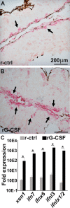
Tadpoles injected with rCSF3 possess more esterase-positive myeloid cells in their kidneys, concurrent with greater Xen1 and IFN gene expression. Tadpoles (NF 54) were injected i.p. with rCSF3 (2 μg/animal) or the recombinant control (r-ctrl), and 24 h later, their kidneys were examined for the proportions of esterase-positive cells (A and B) and Xen1 and IFN gene expression (C). Images in A and B are representative of results derived from 6 individual animals per treatment group (n = 6). The results in C are means + SEM, n = 6. Gene expression (C) was assessed relative to the gapdh endogenous control. Xen1 and IFN gene expression was normalized against the baseline (r-ctrl) expression for the respective genes. Arrows in (A) and (B) indicate esterase-positive cells. Asterisks (*) above lines indicate statistical differences between treatment groups indicated by those lines.
FIG 5
FIG 5
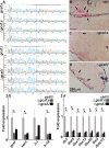
F0 transgenic tadpoles bearing CRISPR-Cas9-induced mutations in csf3.s or csf3.l possess fewer esterase positive myeloid cells in their kidneys, concurrent with lower Xen1 and IFN gene expression therein. (A) Sequenced csf3 loci from cohort controls and CRISPR-Cas9-altered F0 tadpoles. The underlined sequences indicate the guide RNA sequences, the red underlines denote the protospacer adjacent motif (PAM) sites, and the vertical dotted lines represent the cut sites. Esterase-positive cells in the kidneys of cohort controls (B), Δcsf3.s (C), and Δcsf3.s (D) F0 tadpoles. Images in C and D are representative of 3 and 4 tadpoles, respectively. Analysis of CSF3, Xen1, EVR3 to 1, and ERV k-18 (E) and IFN gene expression in cohort controls (N = 6), Δcsf3.s (N = 3), and Δcsf3.s (N = 4) F0 tadpoles (F). The results in E and F are means + SEM. Gene expression (E and F) was assessed relative to the gapdh endogenous control. Arrows in (B-D) indicate esterase-positive cells. Asterisks (*) above lines indicate statistical differences between treatment groups indicated by those lines.

Similar articles

Cited by

References

    1. Chinchar VG, Hyatt A, Miyazaki T, Williams T. 2009. Family Iridoviridae: poor viral relations no longer. Curr Top Microbiol Immunol 328:123–170. 10.1007/978-3-540-68618-7_4. - DOI - PubMed
    1. Williams T, Barbosa-Solomieu V, Chinchar VG. 2005. A decade of advances in iridovirus research. Adv Virus Res 65:173–248. 10.1016/S0065-3527(05)65006-3. - DOI - PubMed
    1. Hoverman JT, Gray MJ, Miller DL. 2010. Anuran susceptibilities to ranaviruses: role of species identity, exposure route, and a novel virus isolate. Dis Aquat Organ 89:97–107. 10.3354/dao02200. - DOI - PubMed
    1. Landsberg JH, Kiryu Y, Tabuchi M, Waltzek TB, Enge KM, Reintjes-Tolen S, Preston A, Pessier AP. 2013. Co-infection by alveolate parasites and frog virus 3-like ranavirus during an amphibian larval mortality event in Florida, USA. Dis Aquat Organ 105:89–99. 10.3354/dao02625. - DOI - PubMed
    1. Reeve BC, Crespi EJ, Whipps CM, Brunner JL. 2013. Natural stressors and ranavirus susceptibility in larval wood frogs (Rana sylvatica). Ecohealth 10:190–200. 10.1007/s10393-013-0834-6. - DOI - PubMed

Publication types

LinkOut - more resources