. 2022 Jun 8;96(11):e0010922.
doi: 10.1128/jvi.00109-22. Epub 2022 May 16.

Diversity and Long-Term Dynamics of Human Blood Anelloviruses

Affiliations

Diversity and Long-Term Dynamics of Human Blood Anelloviruses

Joanna Kaczorowska et al. J Virol. .

Abstract

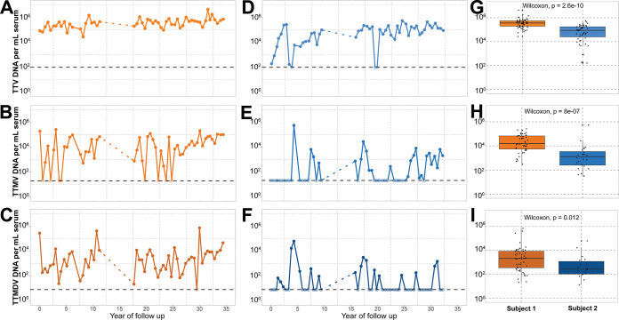
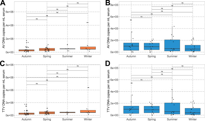
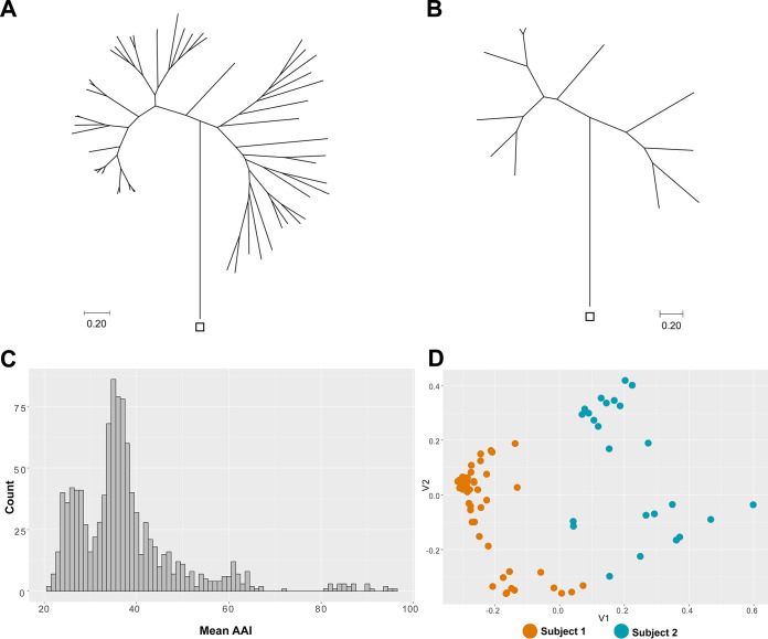
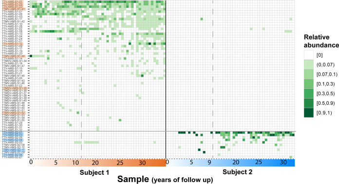
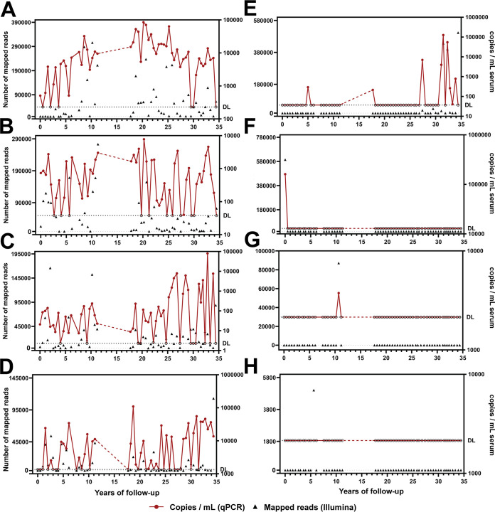
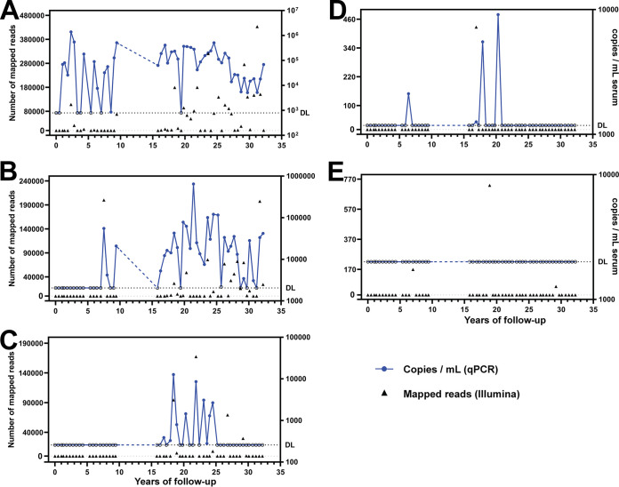
Anelloviruses (AVs) are commensal members of the human blood virome. Even though it was estimated that over 90% of the human population carries AVs, the dynamics of the AV virome ("anellome") are unknown. We investigated the dynamics of blood anellomes in two healthy people followed up for more than 30 years. Both subjects were positive for AVs in the majority of samples. Alphatorquevirus (torque teno virus [TTV]) was the most common genus in both subjects, followed by Betatorquevirus (torque teno minivirus [TTMV]) and Gammatorquevirus (torque teno midivirus [TTMDV]). Almost five times more lineages were found in subject 1 than in subject 2, and the anellomes differed phylogenetically. Both anellomes remained compositionally stable, and 9 out of 64 AV lineages were detected in over half of the time points. We confirmed the long-term and short-term persistence of 13 lineages by specific quantitative PCR (qPCR). AV lineages were detected in blood for over 30 years. Noticeable differences in anellome richness were found between the tested subjects, but both anellomes remained compositionally stable over time. These findings demonstrate that the human blood anellome is personal and that AV infection is chronic and potentially commensal. IMPORTANCE Knowledge of the persistence of AVs in humans is crucial to our understanding of the nature of AV infection (chronic or acute) and the role of AV in the host. We therefore investigated the dynamics of anellovirus infection in two healthy people followed up for 30 years. Our findings suggest that the human blood anellovirus virome (anellome) remains stable and personal for decades.

Keywords: anellome; anellovirus; blood virome; chronic viral infection; torque teno virus; virome.


Conflict of interest statement

The authors declare no conflict of interest.

Figures

FIG 1
FIG 1
Prevalence of anelloviruses over time. DNA concentrations were measured by qPCRs targeting human-infecting anellovirus genera. (A to F) qPCRs detecting Alphatorquevirus (A), Betatorquevirus (B), and Gammatorquevirus (C) in subject 1 samples and Alphatorquevirus (D), Betatorquevirus (E), and Gammatorquevirus (F) in subject 2 samples. The estimated detection limit of each assay is shown as a black horizontal dashed line, and the samples negative in the assay are depicted as open circles. (G to I) Box plots showing summarized Alphatorquevirus (G), Betatorquevirus (H), and Gammatorquevirus (I) DNA copy numbers in both subjects. The horizontal bar within the box plots depicts the median concentration. The P values are shown at the top of each box plot.
FIG 2
FIG 2
Seasonality of anellovirus infection. (A and B) Subject 1 (A) and subject 2 (B) total AV copy numbers were calculated by summing up the results of all three genus-specific qPCRs. (C and D) Copy numbers of TTV in subject 1 (C) and subject 2 (D). ns, not significant.
FIG 3
FIG 3
Highly diverse and personal anellome. (A and B) Phylogenetic relationships of 53 subject 1 (A) and 11 subject 2 (B) anellovirus lineages. The trees were constructed using the maximum likelihood method and are based on amino acid alignments of ORF1 proteins. The outgroup (rodent anellovirus [GenBank accession number NC_040687]) is indicated with a square. (C) Pairwise amino acid identity (AAI) comparison between anellovirus lineages derived from both subjects. All ORF1 sequences were compared against each other. (D) Principal-coordinate analysis (PCoA) ordination of the viral community matrix based on the unweighted UniFrac divergence metric. The color code of the subjects is given at the bottom.
FIG 4
FIG 4
Anellovirus diversity and prevalence over time. A heatmap shows the relative abundances of various anellovirus lineages in subject 1 and subject 2 over time. The color strength represents the relative abundance of a lineage per time point. The approximate years of follow-up are shown on the x axis. The lineages were arranged on the y axis based on the number of positive subject time points. The lineages highlighted with orange or blue were also tested by specific qPCR. The dashed lines denote sampling gaps.
FIG 5
FIG 5
Persistence of selected anellovirus lineages over time in subject 1. Eight lineages were selected for specific qPCR analysis. The graphs show the results of mapping of the Illumina reads to the selected lineage (left y axis) and the copies per milliliter measured using lineage-specific qPCR (right y axis). The analysis was performed for the following lineages: TTV-AMS-S1-01 (A), TTV-AMS-S1-23 (B), TTV-AMS-S1-25 (C), TTMV-AMS-S1-41 (D), TTV-AMS-S1-12 (E), TTMV-AMS-S1-44 (F), TTMV-AMS-S1-52 (G), and TTMDV-AMS-S1-35 (H). One out of two performed qPCR runs is plotted here. The dashed horizontal line marked with “DL” represents the estimated limit of detection of the qPCR assay. The samples that were negative by qPCR are depicted with an open circle.
FIG 6
FIG 6
Persistence of selected anellovirus lineages over years in subject 2. Five lineages were selected for specific qPCR analysis. The graphs show the results of mapping of the Illumina reads to the selected lineage (left y axis) and the copies per milliliter measured using lineage-specific qPCR (right y axis). The analysis was performed for the following lineages: TTV-AMS-S2-01 (A), TTV-AMS-S2-03 (B), TTV-AMS-S2-04 (C), TTV-AMS-S2-06 (D), and TTMV-AMS-S2-11 (E). One out of two performed qPCR runs is plotted here. The dashed horizontal line marked with “DL” represents the estimated limit of detection of the qPCR assay. The samples that were negative by qPCR are depicted with an open circle.

Similar articles

Cited by

References

    1. Moustafa A, Xie C, Kirkness E, Biggs W, Wong E, Turpaz Y, Bloom K, Delwart E, Nelson KE, Venter JC, Telenti A. 2017. The blood DNA virome in 8,000 humans. PLoS Pathog 13:e1006292. 10.1371/journal.ppat.1006292. - DOI - PMC - PubMed
    1. Vu D-L, Cordey S, Simonetta F, Brito F, Docquier M, Turin L, van Delden C, Boely E, Dantin C, Pradier A, Roosnek E, Chalandon Y, Zdobnov EM, Masouridi-Levrat S, Kaiser L. 2019. Human pegivirus persistence in human blood virome after allogeneic haematopoietic stem-cell transplantation. Clin Microbiol Infect 25:225–232. 10.1016/j.cmi.2018.05.004. - DOI - PubMed
    1. Virgin HW, Wherry EJ, Ahmed R. 2009. Redefining chronic viral infection. Cell 138:30–50. 10.1016/j.cell.2009.06.036. - DOI - PubMed
    1. Kaczorowska J, van der Hoek L. 2020. Human anelloviruses: diverse, omnipresent and commensal members of the virome. FEMS Microbiol Rev 44:305–313. 10.1093/femsre/fuaa007. - DOI - PMC - PubMed
    1. Li G, Zhou Z, Yao L, Xu Y, Wang L, Fan X. 2019. Full annotation of serum virome in Chinese blood donors with elevated alanine aminotransferase levels. Transfusion (Paris) 59:3177–3185. 10.1111/trf.15476. - DOI - PMC - PubMed

Publication types