. 2022 May 17;56(2):248-258.
doi: 10.2478/raon-2022-0016.

The dose accumulation and the impact of deformable image registration on dose reporting parameters in a moving patient undergoing proton radiotherapy

Affiliations

The dose accumulation and the impact of deformable image registration on dose reporting parameters in a moving patient undergoing proton radiotherapy

Gasper Razdevsek et al. Radiol Oncol. .

Abstract

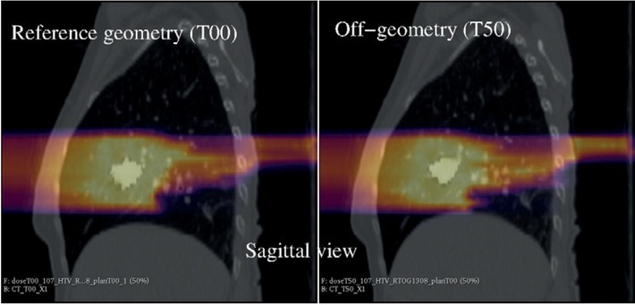
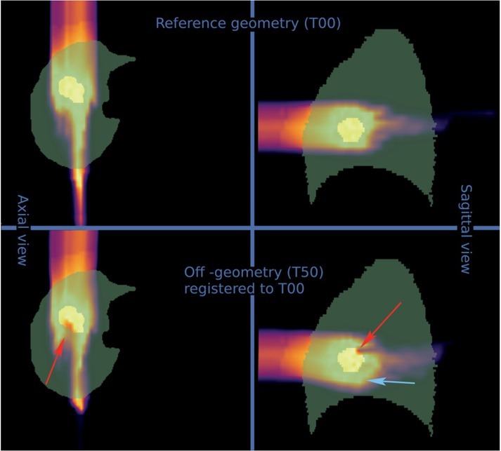
Introduction: Potential changes in patient anatomy during proton radiotherapy may lead to a deviation of the delivered dose. A dose estimate can be computed through a deformable image registration (DIR) driven dose accumulation. The present study evaluates the accumulated dose uncertainties in a patient subject to an inadvertent breathing associated motion.

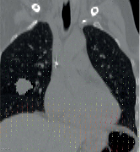
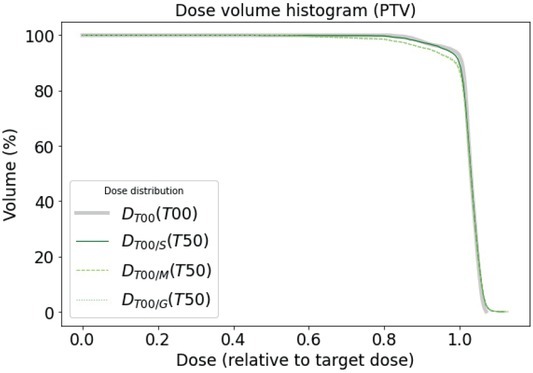
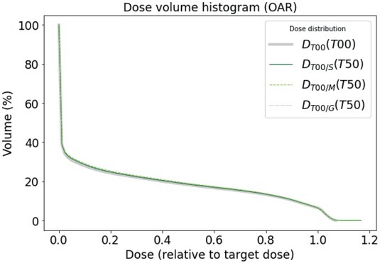
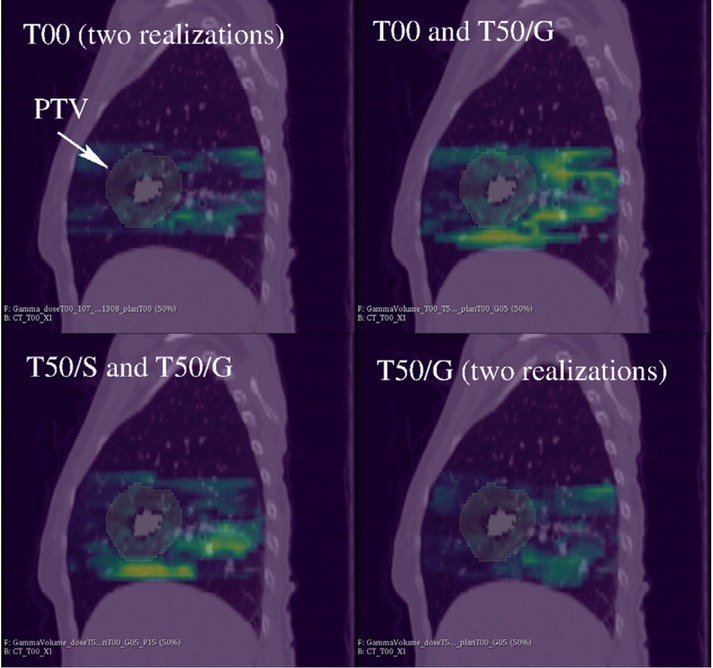
Materials and methods: A virtual lung tumour was inserted into a pair of single participant landmark annotated computed tomography images depicting opposite breathing phases, with the deep inspiration breath-hold the planning reference and the exhale the off-reference geometry. A novel Monte Carlo N-Particle, Version 6 (MCNP6) dose engine was developed, validated and used in treatment plan optimization. Three DIR methods were compared and used to transfer the exhale simulated dose to the reference geometry. Dose conformity and homogeneity measures from International Committee on Radioactivity Units and Measurements (ICRU) reports 78 and 83 were evaluated on simulated dose distributions registered with different DIR algorithms.

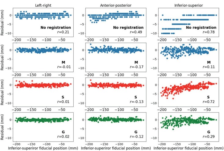
Results: The MCNP6 dose engine handled patient-like geometries in reasonable dose calculation times. All registration methods were able to align image associated landmarks to distances, comparable to voxel sizes. A moderate deterioration of ICRU measures was encountered in comparing doses in on and off-reference anatomy. There were statistically significant DIR driven differences in ICRU measures, particularly a 10% difference in the relative D98% for planning tumour volume and in the 3 mm/3% gamma passing rate.

Conclusions: T he dose accumulation over two anatomies resulted in a DIR driven uncertainty, important in reporting the associated ICRU measures for quality assurance.

Keywords: MCNP6; Monte-Carlo; adaptive therapy; dose distribution measurement; dose homogeneity; image registration; proton therapy.


Figures

Figure 1
Figure 1
A coronal view of the deformable image transformation deformation field of moving exhale phase with respect to the reference deep inspiration breath-hold (DIBH) phase. Size and direction of the local motion is indicated by the arrows with the colour/whiteness of the arrow indicating the size of the translation.
Figure 2
Figure 2
Scatter plots of the annotated landmarks with respect to the residual shift between expiration and inspiration positions on y-axis and location of landmark along the inferior-superior direction on the x-axis. A total of twelve panes indicate residual extent in the left-right direction (first column), anterior-posterior direction (second column) and inferior-superior direction (third column). The top row shows differences in landmark locations without registration, following rows correspond to (S) Staring deformable image registration (DIR), (G) Guy DIR and (M) Mattes DIR algorithms. Indicated numbers correspond to Pearson’s r coefficient calculated for the shown distribution.
Figure 3
Figure 3
A sagittal view of the pair of the 4D CT image corresponding to the extreme breathing frames, left: deep inspiration breath-hold (DIBH) reference geometry, T00, right: exhale off-reference geometry, T50. A tumour delineated in Non-Small Cell Lung Cancer (NSCLC) Radiomics study, case R005, was artificially added into the lung. Dose delivered according to beamlet set that was optimized for irradiation in the reference frame is superimposed with colour scale/grey scale corresponding to the accumulated dose where bright colours/white corresponds to a higher accumulated dose. A slight relative upward motion of tumour in T50 geometry causes the right pane dose distribution to deviate from the planned distribution illustrated in the left pane.
Figure 4
Figure 4
Colormap/grayscale image of dose distributions superimposed on lung mask for two views (axial and sagittal) and two irradiation conditions - irradiation geometry identical to planned geometry, T00, top panes and irradiation geometry non equal to the planned geometry, with dose registered back to the reference geometry T50, below. Red/dark arrows indicate areas of under-treatment for the off-geometry case. Blue/light arrows indicate areas of over-treatment in organs at risk.
Figure 5
Figure 5
Dose-volume histogram (DVH) for planning tumour volume (PTV) under different irradiation conditions and volume representations. Comparison of DT00(T00), thick solid line DVH for irradiation geometry identical to planned geometry, and DT00/S(T50) DT00/M(T50) and DT00/G(T50), solid, dashed and dotted lines for DVH in off-geometry but evaluated in the reference geometry using Staring (S), Mattes (M) or Guy (G) deformable image registration (DIR) method, respectively. Curves for Mattes and Guy registration methods nearly overlap.
Figure 6
Figure 6
Dose-volume histogram (DVH) for planned organ at risk volume of organ at risk (OAR), left lung, under different irradiation conditions and volume representations. Comparison of DT00(T00), thick solid line DVH for irradiation geometry identical to planned geometry, and DT00/S(T50), DT00/M(T50) and DT00/G(T50), solid, dashed and dotted lines for DVH in off-geometry but evaluated in the reference geometry using Staring (S), Mattes (M) or Guy (G) deformable image registration (DIR) method, respectively.
Figure 7
Figure 7
Gamma analysis for 3 mm/3% distance to agreement / dose difference tolerance criteria. All panes show sagittal projection of the CT image overlaid with planning tumour volume (PTV), indicated by arrow, and superimposed gamma function, where yellow/bright colour corresponds to a gamma value of approximately 2. Top left and bottom right pane show dose comparison of statistically independent realizations of the same dose plan, top left for reference geometry, bottom-right for off-reference geometry registered with Guy’s deformable image registration (DIR). Top right is a comparison of T50 dose registered to T00 using Guy’s method and T00 dose. Bottom left is a comparison of T50 dose registered to T00 using two registration methods, S for Staring and G for Guy.

Similar articles

References

    1. Pedroni E, Bacher R, Blattmann H, Bohrinaer T, Coray A, Lomax A. The 200-MeV proton therapy project at the Paul Scherrer Institute: conceptual design and practical realization. Med Phys. 1995;22:37–53. doi: 10.1118/1.597522. et al. - DOI - PubMed
    1. Degiovanni A, Amaldi U. History of hadron therapy accelerators. Phys Medica. 2015;31:322–32. doi: 10.1016/j.ejmp.2015.03.002. - DOI - PubMed
    1. Terasawa T, Dvorak T, Ip S, Raman G, Lau J, Trikalinos TA. Systematic review: charged-particle radiation therapy for cancer. Ann Intern Med. 2009;151:556–65. doi: 10.7326/0003-4819-151-8-200910200-00145. - DOI - PubMed
    1. Verma V, Rwigema J-CM, Malyapa RS, Regine WF, Simone CB. Systematic assessment of clinical outcomes and toxicities of proton radiotherapy for reirradiation. Radiother Oncol. 2017;125:21–30. doi: 10.1016/j.radonc.2017.08.005. - DOI - PubMed
    1. Liao Z, Lee JJ, Komaki R, Gomez DR, O’Reilly MS, Fossella FV. Bayesian adaptive randomization trial of passive scattering proton therapy and intensity-modulated photon radiotherapy for locally advanced non–small-cell lung cancer. J Clin Oncol. 2018;36:1813–22. doi: 10.1200/JCO.2017.74.0720. et al. - DOI - PMC - PubMed

Publication types

MeSH terms