Observational Study
. 2022 Jul 1;79(7):682-692.
doi: 10.1001/jamaneurol.2022.1025.

Association of Brain Atrophy With Disease Progression Independent of Relapse Activity in Patients With Relapsing Multiple Sclerosis

Affiliations
Observational Study

Association of Brain Atrophy With Disease Progression Independent of Relapse Activity in Patients With Relapsing Multiple Sclerosis

Alessandro Cagol et al. JAMA Neurol. .

Abstract

Importance: The mechanisms driving neurodegeneration and brain atrophy in relapsing multiple sclerosis (RMS) are not completely understood.

Objective: To determine whether disability progression independent of relapse activity (PIRA) in patients with RMS is associated with accelerated brain tissue loss.

Design, setting, and participants: In this observational, longitudinal cohort study with median (IQR) follow-up of 3.2 years (2.0-4.9), data were acquired from January 2012 to September 2019 in a consortium of tertiary university and nonuniversity referral hospitals. Patients were included if they had regular clinical follow-up and at least 2 brain magnetic resonance imaging (MRI) scans suitable for volumetric analysis. Data were analyzed between January 2020 and March 2021.

Exposures: According to the clinical evolution during the entire observation, patients were classified as those presenting (1) relapse activity only, (2) PIRA episodes only, (3) mixed activity, or (4) clinical stability.

Main outcomes and measures: Mean difference in annual percentage change (MD-APC) in brain volume/cortical thickness between groups, calculated after propensity score matching. Brain atrophy rates, and their association with the variables of interest, were explored with linear mixed-effect models.

Results: Included were 1904 brain MRI scans from 516 patients with RMS (67.4% female; mean [SD] age, 41.4 [11.1] years; median [IQR] Expanded Disability Status Scale score, 2.0 [1.5-3.0]). Scans with insufficient quality were excluded (n = 19). Radiological inflammatory activity was associated with increased atrophy rates in several brain compartments, while an increased annualized relapse rate was linked to accelerated deep gray matter (GM) volume loss. When compared with clinically stable patients, patients with PIRA had an increased rate of brain volume loss (MD-APC, -0.36; 95% CI, -0.60 to -0.12; P = .02), mainly driven by GM loss in the cerebral cortex. Patients who were relapsing presented increased whole brain atrophy (MD-APC, -0.18; 95% CI, -0.34 to -0.02; P = .04) with respect to clinically stable patients, with accelerated GM loss in both cerebral cortex and deep GM. No differences in brain atrophy rates were measured between patients with PIRA and those presenting relapse activity.

Conclusions and relevance: Our study shows that patients with RMS and PIRA exhibit accelerated brain atrophy, especially in the cerebral cortex. These results point to the need to recognize the insidious manifestations of PIRA in clinical practice and to further evaluate treatment strategies for patients with PIRA in clinical trials.


Conflict of interest statement

Conflict of Interest Disclosures: Dr Weigel reported personal fees from the Swiss National Science Fund for magnetic resonance imaging (MRI) protocol development and acquisition during the conduct of the study and grants from Biogen for development of spinal cord MRI for patients with spinal muscular atrophy outside the submitted work. Dr Yaldizli reported grants from ECTRIMS/MAGNIMS, University of Basel, Pro Patient Stiftung University Hospital Basel, Free Academy Basel, and Swiss MS Society and reported advisory board/lecture and consultancy fees from Roche, Sanofi Genzyme, Almirall, Biogen, and Novartis. Dr Oechtering reported grants from Swiss MS Society outside the submitted work. Dr Lorscheider reported grants and other fees from Innosuisse – Swiss Innovation Agency, Novartis, Biogen, Roche, and Teva outside the submitted work. Dr Fischer-Barnicol reported personal fees from Biogen outside the submitted work. Dr Müller reported honoraria or grants from Almirall, Biogen, Celgene, Novartis, Teva, Merck Serono, Genzyme, Roche, and Bayer Schweiz. Dr Achtnichts reported grants from Swiss MS Society during the conduct of the study and personal fees or grants from Swiss MS Society, Celgene, Biogen, Novartis, and Merck outside the submitted work. Dr Vehoff reported personal fees or grants from Roche, Almirall, Biogen, Novartis, Teva, Merck-Serono, Genzyme, and Bayer Schweiz outside the submitted work. Dr Chan reported honoraria from Actelion-Janssen, Almirall, Bayer, Biogen, Celgene, Sanofi-Genzyme, Merck, Novartis, Roche, and Teva, all for hospital research funds; reported research support from Biogen, Genzyme, Roche, UCB, the European Union, and the Swiss National Foundation; and reported being associate editor of European Journal of Neurology, on the editorial board for Clinical and Translational Neuroscience, and topic editor for the Journal of International Medical Research. Dr Salmen reported personal fees from Almirall Hermal, Bristol Myers Squibb, Novartis, Biogen, Merck, Novartis, Roche, and Sanofi Genzyme; grants from Baasch Medicus Foundation and the Swiss MS Society outside the submitted work; and serving on the editorial board of Frontiers in Neurology – Multiple Sclerosis and Neuroimmunology. Dr Zecca reported grants from Biogen, Bristol Myers Squibb, Merck Serono, Teva, Sanofi, Almirall, Lundbeck, Novartis, Roche, Genzyme, Celgene, and Bayer outside the submitted work. Dr Derfuss reported fees from and/or serving on advisory boards or steering committees for Actelion, Alexion, Biogen, Celgene, GeNeuro, MedDay, Merck, Mitsubishi Pharma, Novartis, Roche and Sanofi-Genzyme and reported research support from Alexion, Biogen, Novartis, Roche, Swiss National Research Foundation, University of Basel, and Swiss MS Society. Dr Lalive reported honoraria for speaking from Biogen-Idec, CSL Bering, Merck Serono, Novartis, Sanofi-Aventis, and Teva; consulting fees from Biogen-Idec, Geneuro, Genzyme, Merck Serono, Novartis, Sanofi-Aventis, and Teva; and research grants from Biogen-Idec, Merck Serono, and Novartis. Dr Gobbi reported grants from Bayer, Biogen, Bristol Myers Squibb, Novartis, Teva, Roche, Sanofi, Lundbeck, Genzyme, Celgene, Novartis, and Merck Serono outside the submitted work. Dr Kappos reported grants and other fees from AbbVie, Actelion, Allergan, Almirall AurigaVision, Bayer, Biogen, Bristol Myers Squibb, Celgene, CSL Behring, Desitin, Eisai, EMD Serono, European Union, Genentech, Genzyme, GlaxoSmithKline, InnoSuisse, Janssen, Japan Tobacco Inc, Merck, Minoryx, Neurostatus, Novartis, Pfizer, Roche, Sanofi, Santhera, Senda Biosciences, Shionogi, Shire, TG Therapeutics, Swiss MS Society, and Swiss National Research Foundation. Dr Kuhle reported grants and other support from Swiss MS Society, Swiss National Research Foundation, University of Basel, Progressive MS Alliance, Bayer, Biogen, Bristol Myers Squibb, Celgene, Merck, Novartis, Octave Bioscience, Roche, and Sanofi. Dr Granziera reported that her institution received research support and other fees from Actelion, Genzyme-Sanofi, Novartis, GeNeuro, Roche, and Siemens. No other disclosures were reported.

Figures

Figure 1.
Figure 1.. Study Design
BPF indicates brain parenchymal fraction; EDSS, Expanded Disability Status Scale; PIRA, progression independent of relapse activity; T2LV, T2 lesion volume; WMLs, white matter lesions.
Figure 2.
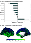
Figure 2.. Annual Percentage Change in Volume and Regional Cortical Thickness
GM indicates gray matter; TBV, total brain volume; WM, white matter.
Figure 3.
Figure 3.. Differences in Rates of Regional Cortical Thinning Comparing Stable Patients vs Patients With Only Progression Independent of Relapse Activity (PIRA) or Patients With Only Relapse Activity
The effect size, expressed as mean difference in annual percentage cortical thickness change (MD-APC), is graphically displayed in different shades of blue for each of the Desikan-Killiany atlas regions presenting significant differences between groups after correction for multiple comparisons.

Similar articles

Cited by

References

    1. Stadelmann C, Wegner C, Brück W. Inflammation, demyelination, and degeneration: recent insights from MS pathology. Biochim Biophys Acta. 2011;1812(2):275-282. doi:10.1016/j.bbadis.2010.07.007 - DOI - PubMed
    1. Lublin FD, Baier M, Cutter G. Effect of relapses on development of residual deficit in multiple sclerosis. Neurology. 2003;61(11):1528-1532. doi:10.1212/01.WNL.0000096175.39831.21 - DOI - PubMed
    1. Lublin FD, Reingold SC, Cohen JA, et al. . Defining the clinical course of multiple sclerosis: the 2013 revisions. Neurology. 2014;83(3):278-286. doi:10.1212/WNL.0000000000000560 - DOI - PMC - PubMed
    1. Kappos L, Wolinsky JS, Giovannoni G, et al. . Contribution of relapse-independent progression vs relapse-associated worsening to overall confirmed disability accumulation in typical relapsing multiple sclerosis in a pooled analysis of 2 randomized clinical trials. JAMA Neurol. 2020;77(9):1132-1140. doi:10.1001/jamaneurol.2020.1568 - DOI - PMC - PubMed
    1. Kappos L, Butzkueven H, Wiendl H, et al. ; Tysabri® Observational Program (TOP) Investigators . Greater sensitivity to multiple sclerosis disability worsening and progression events using a roving versus a fixed reference value in a prospective cohort study. Mult Scler. 2018;24(7):963-973. doi:10.1177/1352458517709619 - DOI - PMC - PubMed

Publication types

MeSH terms