

Recurrent KAT6B/A::KANSL1 Fusions Characterize a Potentially Aggressive Uterine Sarcoma Morphologically Overlapping With Low-grade Endometrial Stromal Sarcoma
- PMID: 35575789
- PMCID: PMC9388494
- DOI: 10.1097/PAS.0000000000001915
Recurrent KAT6B/A::KANSL1 Fusions Characterize a Potentially Aggressive Uterine Sarcoma Morphologically Overlapping With Low-grade Endometrial Stromal Sarcoma
Abstract
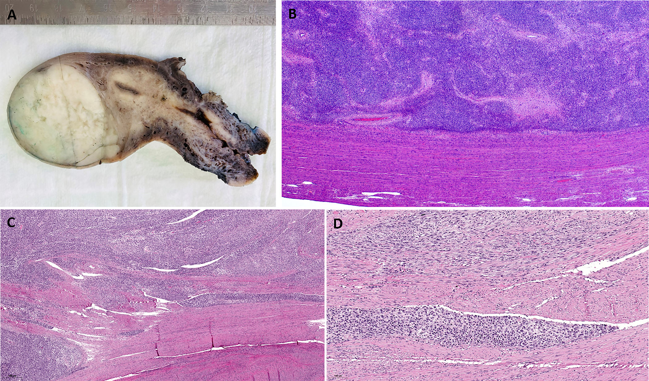
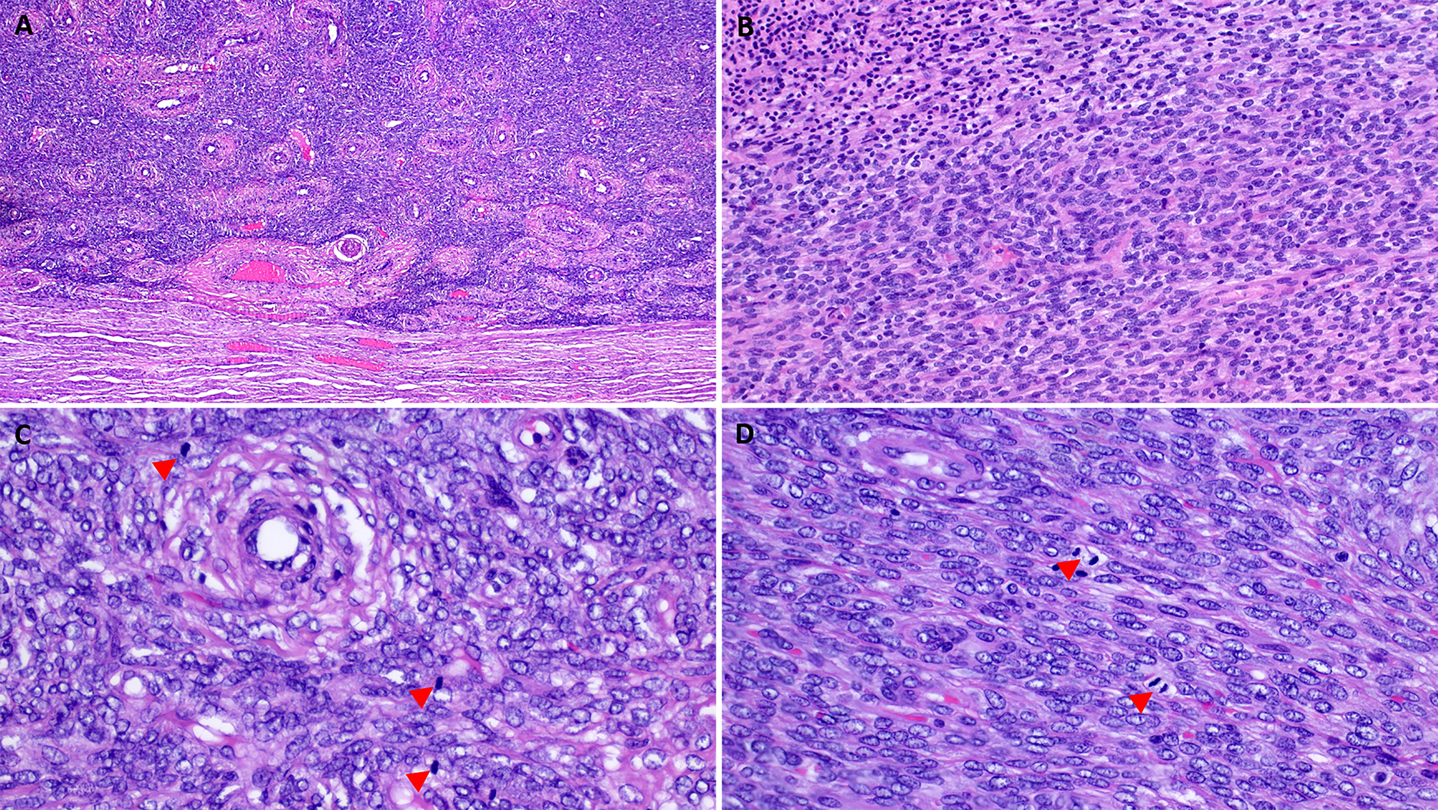
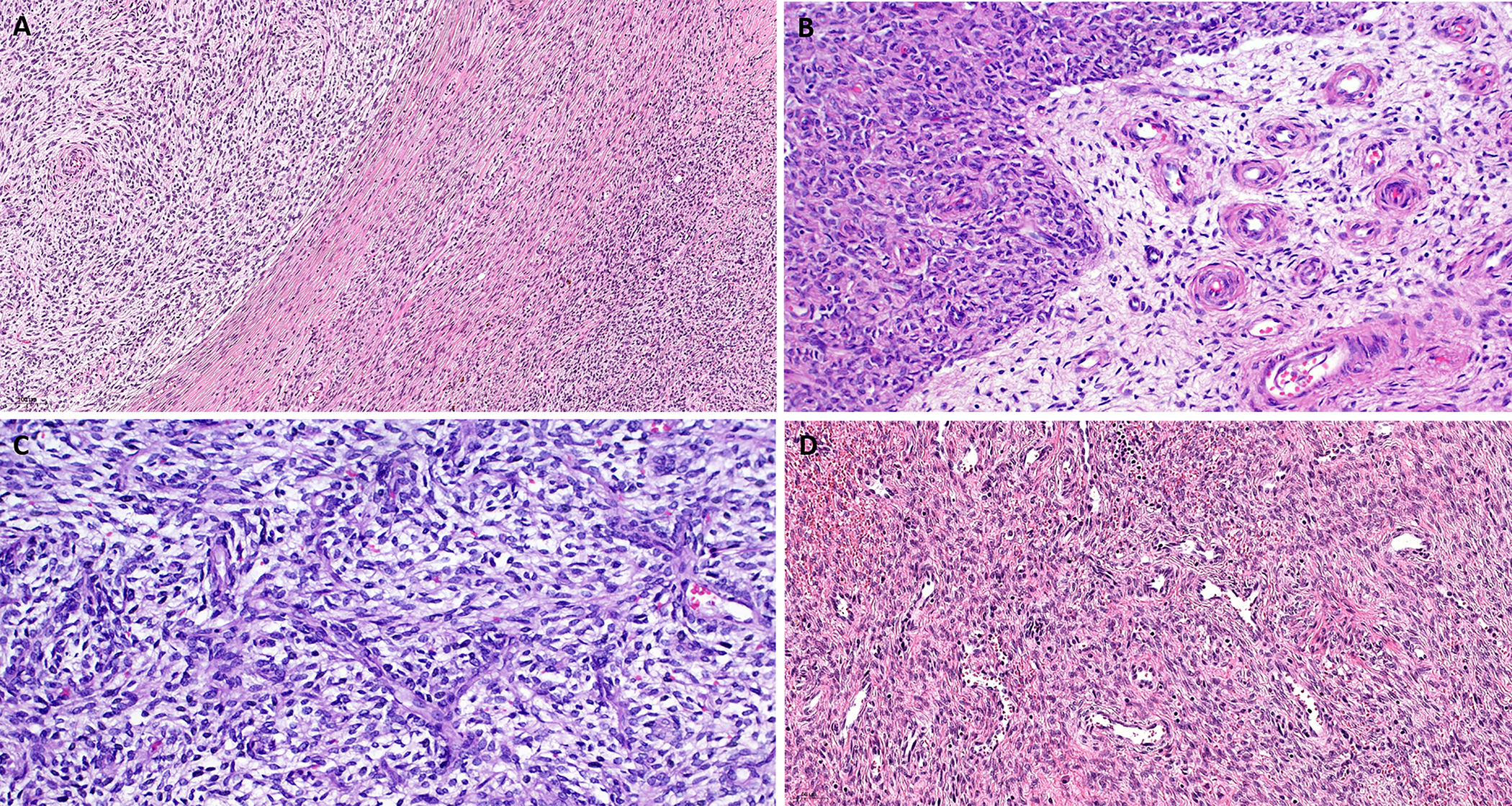
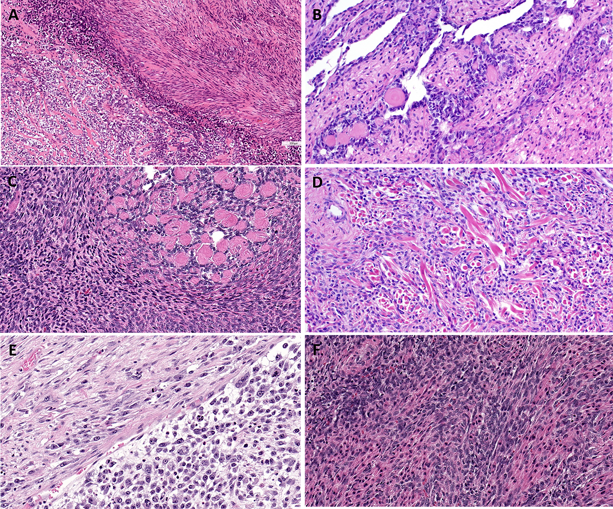
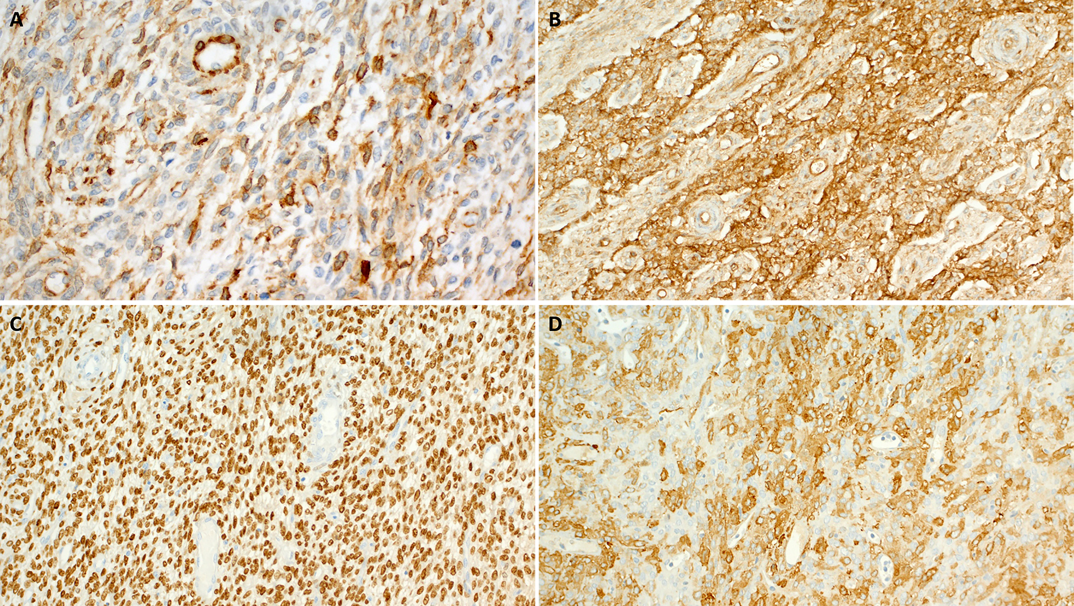
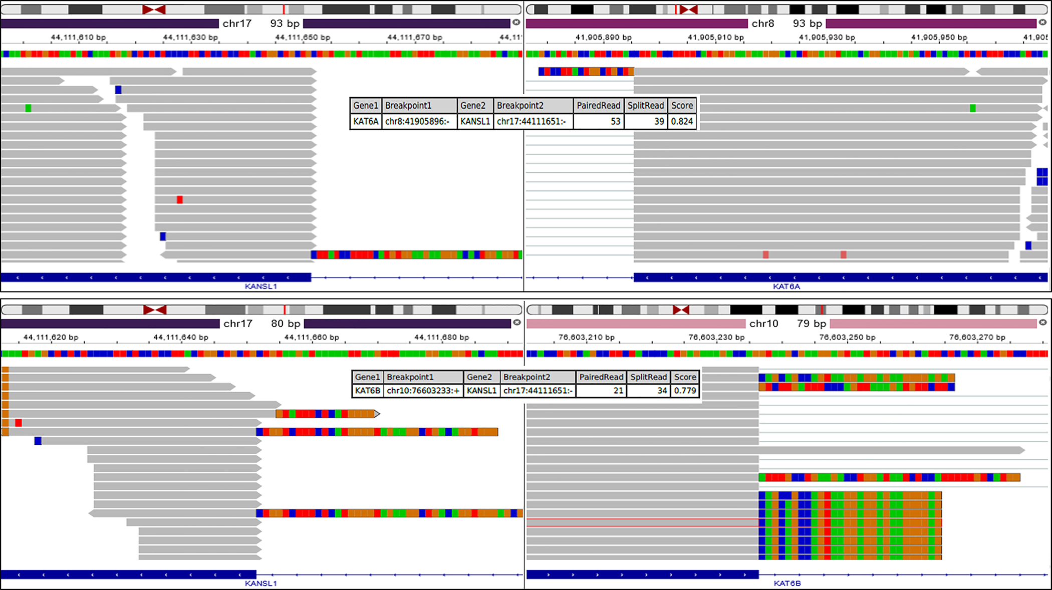
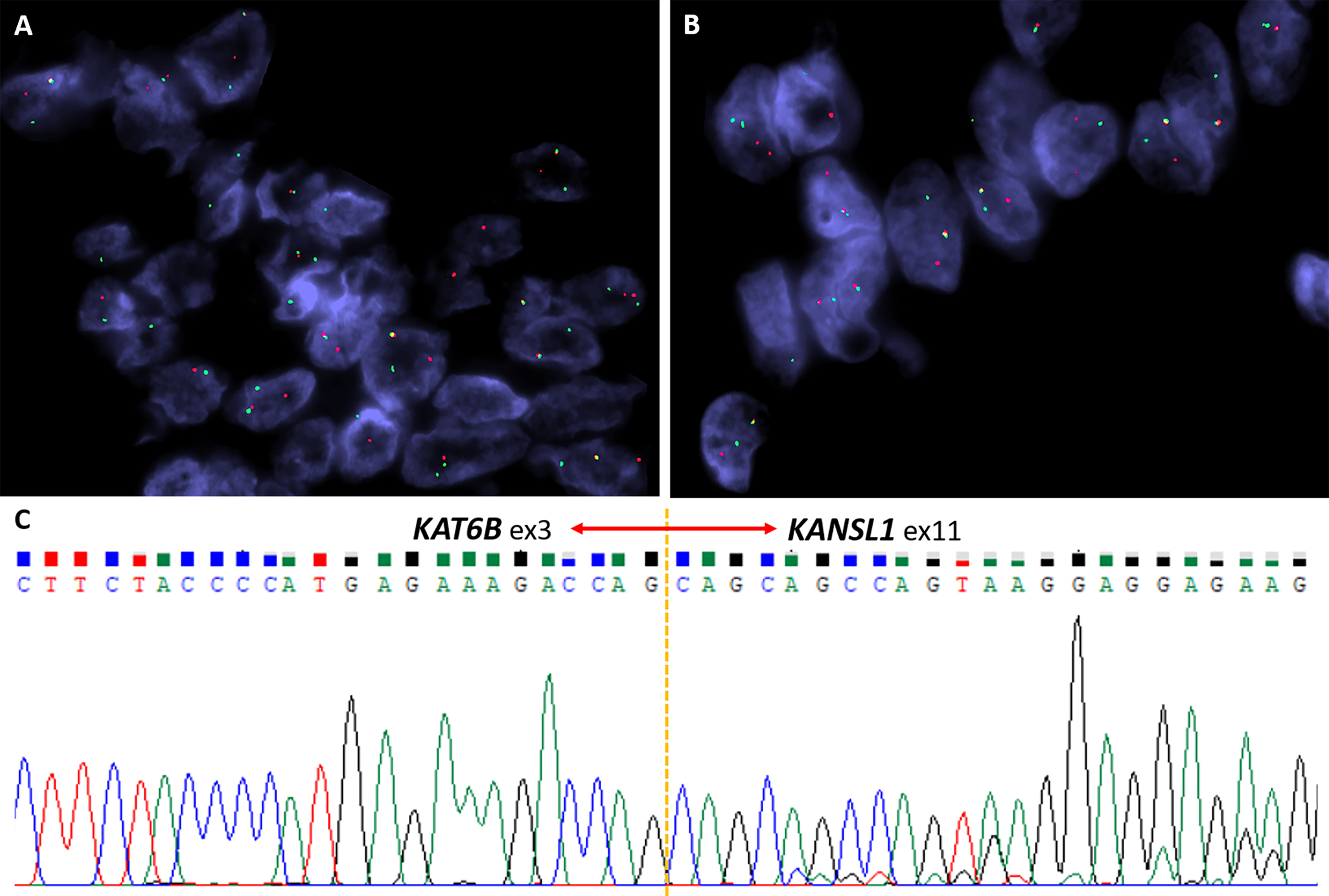
With the widespread application of next-generation sequencing, the genetic landscape of uterine mesenchymal neoplasms has been evolving rapidly to include several recently identified fusion genes. Although chromosomal rearrangements involving the 10q22 and 17q21.31 loci have been reported in occasional uterine leiomyomas decades ago, the corresponding KAT6B::KANSL1 fusion has been only recently identified in 2 uterine tumors diagnosed as leiomyoma and leiomyosarcoma. We herein describe 13 uterine stromal neoplasms carrying a KAT6B::KANSL1 (n=11) and KAT6A::KANSL1 (n=2) fusion. Patient ages ranged from 33 to 81 years (median, 49 y). Tumor size was 2.6 to 23.5 cm (median, 8.2 cm). Nine tumors were myometrium-centered, and 3 had an intracavitary component. Original diagnoses were mostly low-grade endometrial stromal sarcoma (LG-ESS; 10 cases) with atypical features (limited CD10 expression, sex cord-like features, pericytic vasculature, and frequent myxoid changes). Treatment was hysterectomy±bilateral salpingo-oophorectomy (10), myomectomy (1), and curettage (2). Five patients were disease-free at 6 to 34 months, 3 (27%) died of disease at 2 to 47 months, and 3 were alive with disease at 2, 17, and 17 years. Histologically, most tumors showed variable overlap with LG-ESS, but they were generally well-circumscribed lacking the extensive permeative and angioinvasive growth typical of LG-ESS. They were composed of monotonous medium-sized oval and spindle cells arranged into diffuse sheets with prominent spiral-type arterioles and frequent pericytoma-like vascular pattern. Variable myxoid stromal changes were frequent. Mitotic activity ranged from 1 to >20 in 10 HPFs. Immunohistochemistry showed variable expression of CD10 (12/13), estrogen receptor (8/11), progesterone receptor (8/11), smooth muscle actin (9/11), desmin (4/12), h-caldesmon (2/10), calretinin (3/8), inhibin (1/7), WT1 (4/7), cyclin D1 (5/11; diffuse in only 1 case), and pankeratin (5/10). This series characterizes a KAT6B/A::KANSL1 fusion-positive uterine stromal neoplasm within the morphologic spectrum of LG-ESS but with atypical features. The relationship of these neoplasms to genuine LG-ESS remains unclear. This molecular subtype of uterine endometrial stromal sarcoma has the potential for an unfavorable clinical course despite the absence of widely invasive growth; nevertheless, analysis of more cases is necessary to delineate the phenotypic spectrum and biological potential of this tumor.
Copyright © 2022 Wolters Kluwer Health, Inc. All rights reserved.
Conflict of interest statement
Conflicts of Interest and Source of Funding: Supported by P50 CA 140146-01 (to C.R.A.), P50 CA217694 (to C.R.A.), P30 CA008748 (to C.R.A.). This study was supported in part by the research fund from the Ministry of Science and Technology, Taiwan (MOST 109-2326-B-002-010-MY3) to J.-C.L. The authors have disclosed that they have no significant relationships with, or financial interest in, any commercial companies pertaining to this article.
Figures
References
-
- Rommel B, Holzmann C, Bullerdiek J. Malignant mesenchymal tumors of the uterus - time to advocate a genetic classification. Expert Rev Anticancer Ther 2016;16:1155–1166. - PubMed
-
- Ferreira J, Félix A, Lennerz JK, Oliva E. Recent advances in the histological and molecular classification of endometrial stromal neoplasms. Virchows Arch 2018;473:665–678. - PubMed
-
- Momeni-Boroujeni A, Chiang S. Uterine mesenchymal tumours: recent advances. Histopathology 2020;76:64–75. - PubMed
-
- Chiang S Recent advances in smooth muscle tumors with PGR and PLAG1 gene fusions and myofibroblastic uterine neoplasms. Genes Chromosomes Cancer 2021;60:138–146. - PubMed
-
- Arias-Stella JA 3rd, Benayed R, Oliva E, Young RH, Hoang LN, Lee CH, Jungbluth AA, Frosina D, Soslow RA, Antonescu CR, Ladanyi M, Chiang S. Novel PLAG1 Gene Rearrangement Distinguishes a Subset of Uterine Myxoid Leiomyosarcoma From Other Uterine Myxoid Mesenchymal Tumors. Am J Surg Pathol 2019;43:382–388. - PMC - PubMed
Publication types
MeSH terms
Substances
Grants and funding
LinkOut - more resources
Full Text Sources
Medical
Molecular Biology Databases
Research Materials
