

Segmenting electroencephalography wires reduces radiofrequency shielding artifacts in simultaneous electroencephalography and functional magnetic resonance imaging at 7 T
- PMID: 35575944
- PMCID: PMC9323442
- DOI: 10.1002/mrm.29298
Segmenting electroencephalography wires reduces radiofrequency shielding artifacts in simultaneous electroencephalography and functional magnetic resonance imaging at 7 T
Abstract
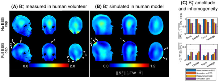
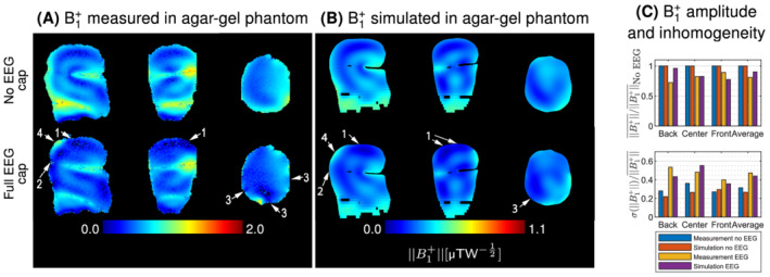
Purpose: Simultaneous scalp electroencephalography and functional magnetic resonance imaging (EEG-fMRI) enable noninvasive assessment of brain function with high spatial and temporal resolution. However, at ultra-high field, the data quality of both modalities is degraded by mutual interactions. Here, we thoroughly investigated the radiofrequency (RF) shielding artifact of a state-of-the-art EEG-fMRI setup, at 7 T, and design a practical solution to limit this issue.
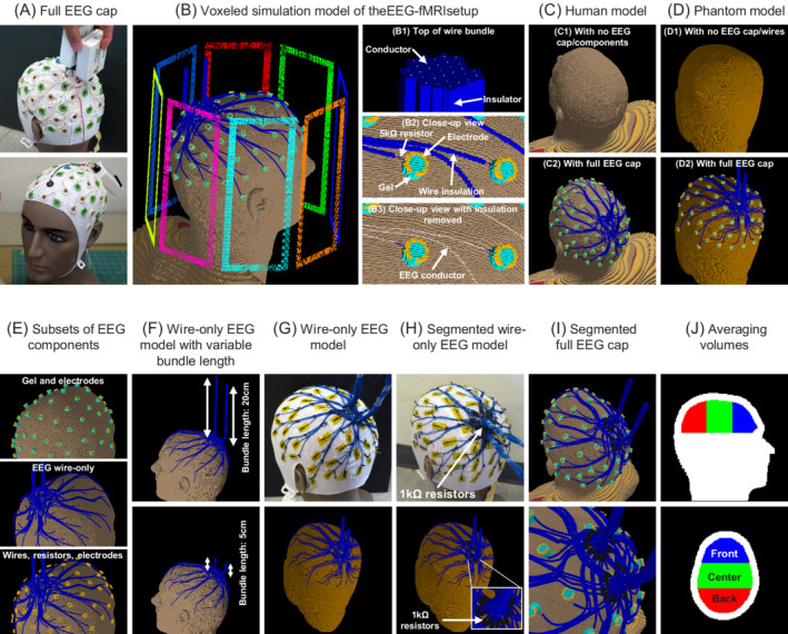
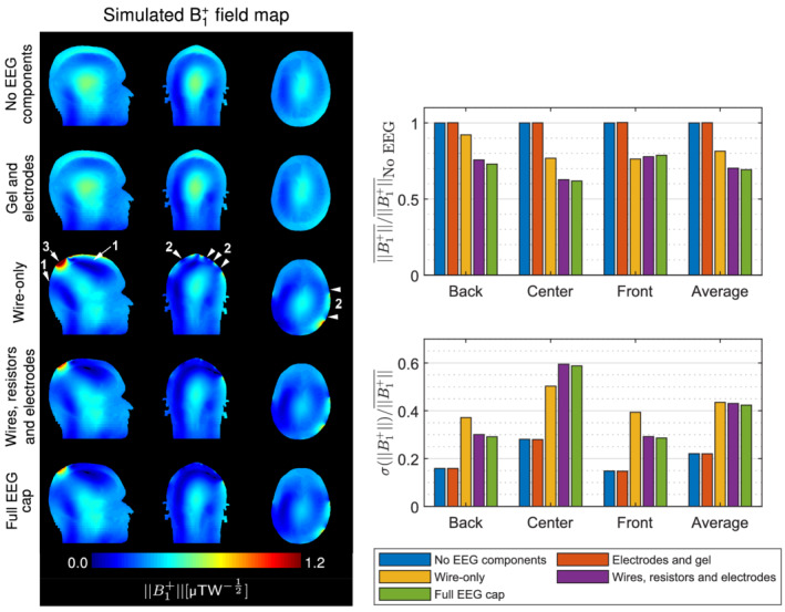
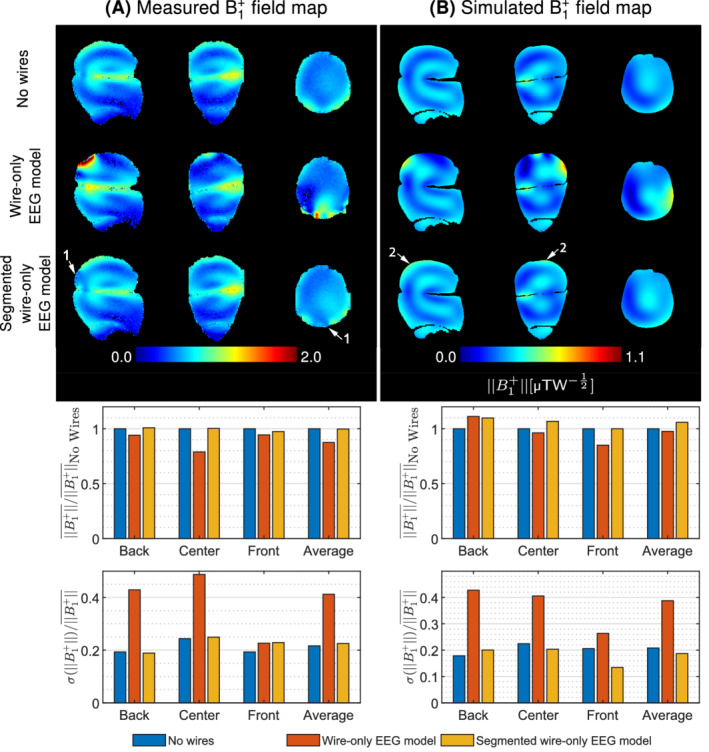
Methods: Electromagnetic field simulations and MR measurements assessed the shielding effect of the EEG setup, more specifically the EEG wiring. The effectiveness of segmenting the wiring with resistors to reduce the transmit field disruption was evaluated on a wire-only EEG model and a simulation model of the EEG cap.
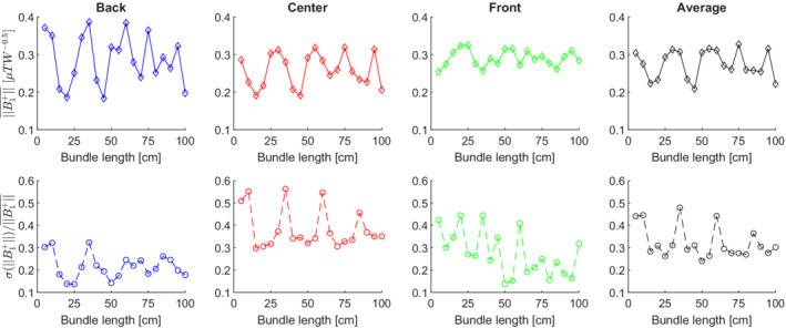
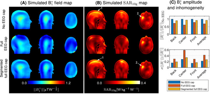
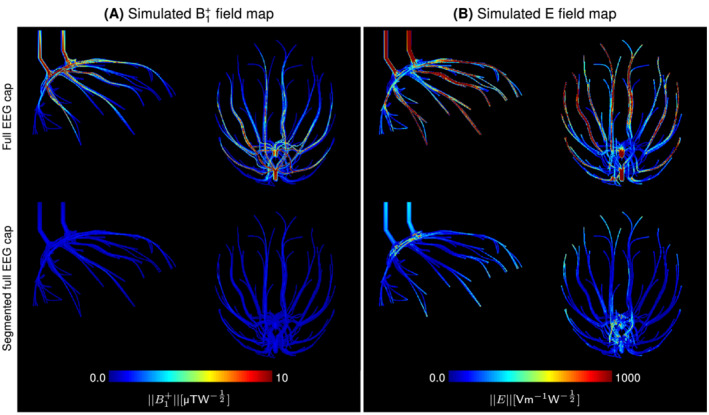
Results: The EEG wiring was found to exert a dominant effect on the disruption of the transmit field, whose intensity varied periodically as a function of the wire length. Breaking the electrical continuity of the EEG wires into segments shorter than one quarter RF wavelength in air (25 cm at 7 T) reduced significantly the RF shielding artifacts. Simulations of the EEG cap with segmented wires indicated similar improvements for a moderate increase of the power deposition.
Conclusion: We demonstrated that segmenting the EEG wiring into shorter lengths using commercially available nonmagnetic resistors is effective at reducing RF shielding artifacts in simultaneous EEG-fMRI. This prevents the formation of RF-induced standing waves, without substantial specific absorption rate (SAR) penalties, and thereby enables benefiting from the functional sensitivity boosts achievable at ultra-high field.
Keywords: 7T; EEG cap; EEG-fMRI; electromagnetic simulations; shielding artifacts; ultra-high field.
© 2022 The Authors. Magnetic Resonance in Medicine published by Wiley Periodicals LLC on behalf of International Society for Magnetic Resonance in Medicine.
Figures
References
-
- Debener S, Ullsperger M, Siegel M, Engel AK. Single‐trial EEG‐fMRI reveals the dynamics of cognitive function. Trends Cogn Sci. 2006;10:558‐563. - PubMed
-
- Herrmann CS, Debener S. Simultaneous recording of EEG and BOLD responses: a historical perspective. Int J Psychophysiol. 2008;67:161‐168. - PubMed
-
- Lemieux L, Salek‐Haddadi A, Josephs O, et al. Event‐related fMRI with simultaneous and continuous EEG: description of the method and initial case report. Neuroimage. 2001;14:780‐787. - PubMed
-
- Rosenkranz K, Lemieux L. Present and future of simultaneous EEG‐fMRI. MAGMA. 2010;23:309‐316. - PubMed
Publication types
MeSH terms
LinkOut - more resources
Full Text Sources
Miscellaneous
