. 2022 May 16;18(5):e1010533.
doi: 10.1371/journal.ppat.1010533. eCollection 2022 May.

Deactivation of the antiviral state by rabies virus through targeting and accumulation of persistently phosphorylated STAT1

Affiliations

Deactivation of the antiviral state by rabies virus through targeting and accumulation of persistently phosphorylated STAT1

Gayathri Manokaran et al. PLoS Pathog. .

Abstract

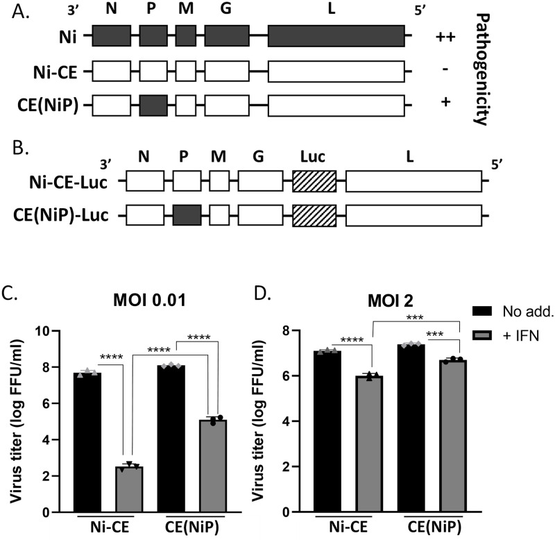
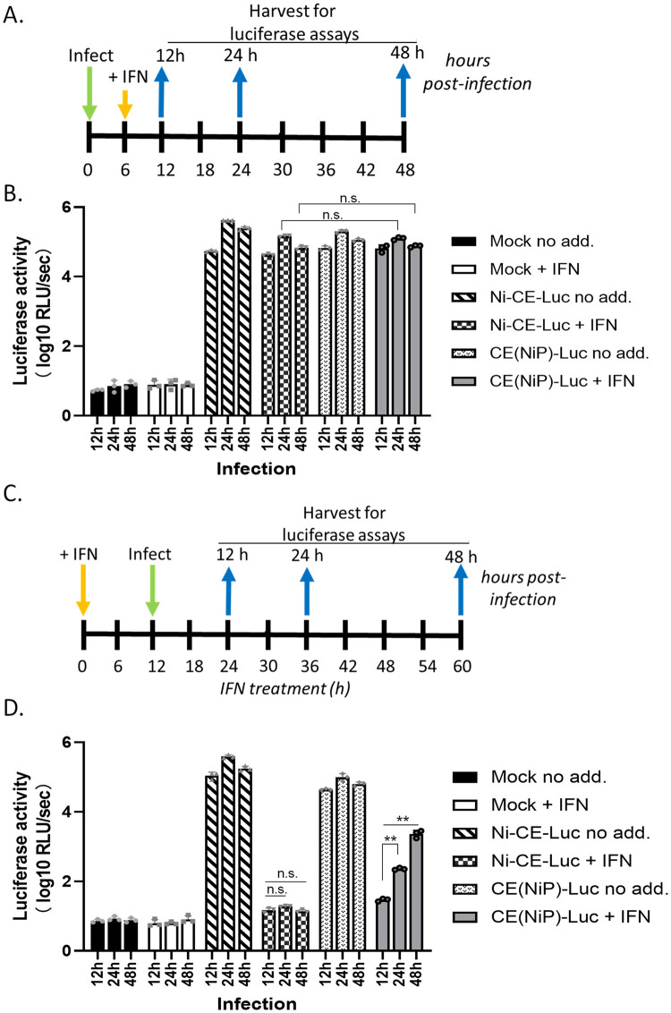
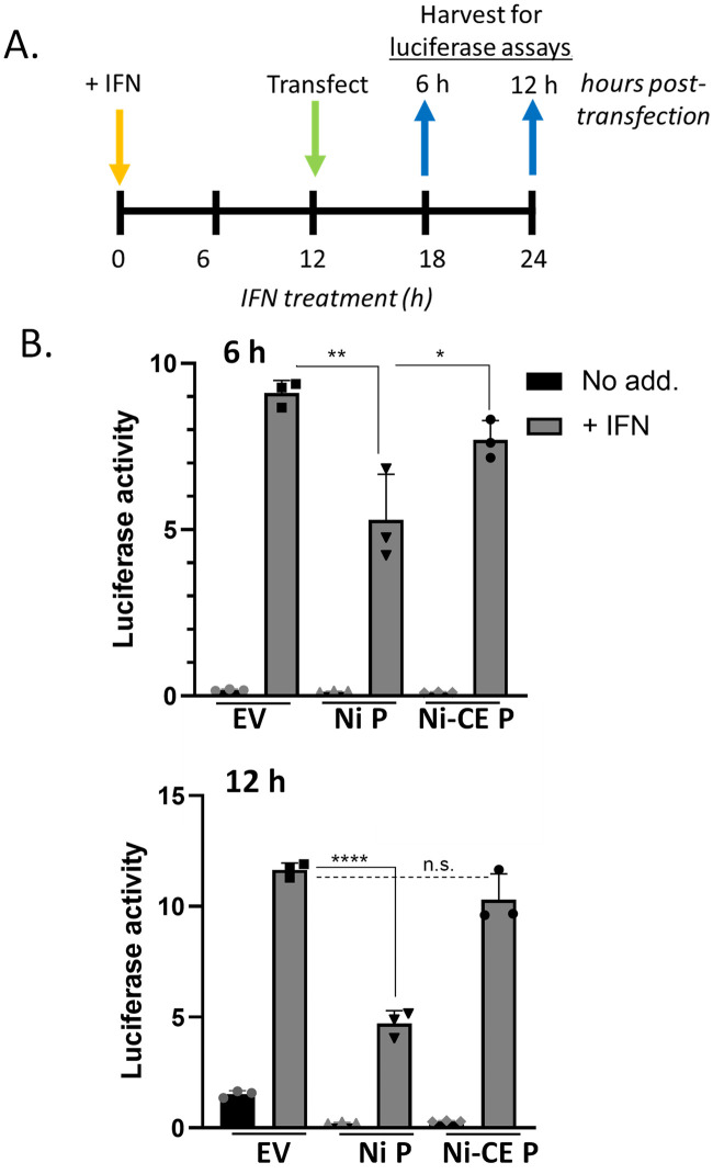
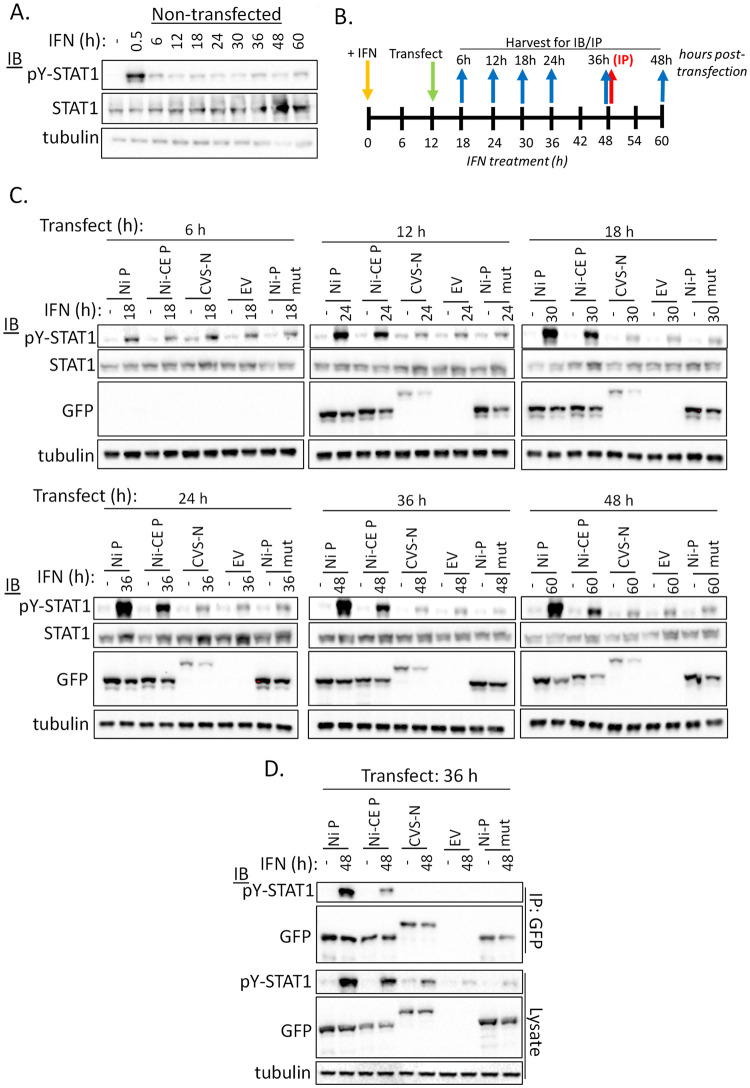
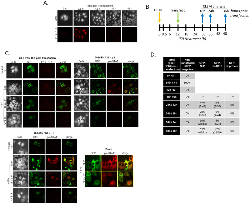
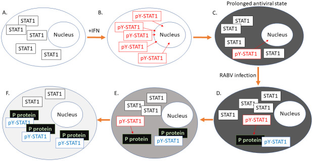
Antagonism of the interferon (IFN)-mediated antiviral state is critical to infection by rabies virus (RABV) and other viruses, and involves interference in the IFN induction and signaling pathways in infected cells, as well as deactivation of the antiviral state in cells previously activated by IFN. The latter is required for viral spread in the host, but the precise mechanisms involved and roles in RABV pathogenesis are poorly defined. Here, we examined the capacity of attenuated and pathogenic strains of RABV that differ only in the IFN-antagonist P protein to overcome an established antiviral state. Importantly, P protein selectively targets IFN-activated phosphorylated STAT1 (pY-STAT1), providing a molecular tool to elucidate specific roles of pY-STAT1. We find that the extended antiviral state is dependent on a low level of pY-STAT1 that appears to persist at a steady state through ongoing phosphorylation/dephosphorylation cycles, following an initial IFN-induced peak. P protein of pathogenic RABV binds and progressively accumulates pY-STAT1 in inactive cytoplasmic complexes, enabling recovery of efficient viral replication over time. Thus, P protein-pY-STAT1 interaction contributes to 'disarming' of the antiviral state. P protein of the attenuated RABV is defective in this respect, such that replication remains suppressed over extended periods in cells pre-activated by IFN. These data provide new insights into the nature of the antiviral state, indicating key roles for residual pY-STAT1 signaling. They also elucidate mechanisms of viral deactivation of antiviral responses, including specialized functions of P protein in selective targeting and accumulation of pY-STAT1.


Conflict of interest statement

I have read the journal’s policy and the authors of this manuscript have the following competing interests: G.W.M. holds a patent application (PCT/AU2019/050908) and Australian provisional patent (No. 201901137); ‘‘Novel Viruses’’ 2019.

Figures

Fig 1
Fig 1. IFN sensitivity of Ni-CE and CE(NiP) viruses is dependent on MOI.
(A,B) Schematic representation of genomes of Ni, Ni-CE and CE(NiP) viruses with relative pathogenicity (A), and of viruses containing the luciferase (Luc) gene (B). (C) SK-N-SH cells infected with the indicated virus at MOI of 0.01 or MOI of 2 were incubated in media containing IFN (+ IFN) or without IFN (No add.) at 500 U/ml for 3 or 2 days, respectively, before collection of supernatant and titration (mean with SD, n = 3). ***, p ≤ 0.001; ****, p ≤ 0.0001.
Fig 2
Fig 2. Ni-CE and CE(NiP) viruses are resistant to IFN signaling in infected cells but differ in replication in cells pre-activated by IFN.
(A) and (C) show schematics of timelines for experiments shown in (B) and (D), respectively. (A, B) SK-N-SH cells were infected with the indicated virus (MOI of 3), or mock infected, for 6 h before incubation in media without (no add.) or with IFN (+ IFN) for the indicated times post-infection, and harvesting of cells for measurement of luciferase activity. (C, D) SK-N-SH cells were incubated with or without IFN for 12 h before infection with the indicated virus (MOI of 3), or mock infection, for the indicated times, and harvesting for measurement of luciferase activity. Results of luciferase assays (B, D) show mean with SD (n = 3). n.s., p > 0.05; **, p ≤ 0.01.
Fig 3
Fig 3. IFN/STAT1 signaling persists for over 12 h and is differentially inhibited by expression of Ni and Ni-CE P proteins.
(A) Timeline of experiments performed in (B); SK-N-SH cells were treated with or without IFN for 12 h before transfection with plasmid to express Ni or Ni-CE P protein, or empty vector control (EV), together with plasmids for the IFN/STAT1-dependent luciferase reporter assay; luciferase activity was measured 6 h or 12 h later. (B) Results of luciferase assays show mean with SD (n = 3). n.s., p > 0.05; *, p ≤ 0.05; **, p ≤ 0.01; ****, p ≤ 0.0001.
Fig 4
Fig 4. Ni-CE P protein is defective for antagonism of persistent IFN/STAT1 signaling.
(A) Timeline of experiments performed in (B); COS-7 cells were transfected with plasmids to express Ni P protein, Ni-CE P protein, or CVS-N protein (control) fused to the C-terminus of GFP, together with plasmids for the IFN/STAT1-dependent luciferase reporter assay, followed by treatment with IFN at 18 h and harvesting of cell samples for luciferase assays at the indicated times. (B) Results of dual luciferase assays, showing mean with SD (n = 3). (C) Timeline of experiments performed in (D) and (E); COS-7 (D) or HEK293T (E) were incubated in media without or with IFN for 12 h before transfection with plasmids to express GFP-fused Ni or Ni-CE P proteins or CVS-N protein, together with plasmids for the luciferase reporter assay; cells were then harvested at the indicated time points. Results of luciferase assays using (D) COS-7 and (E) HEK293T cells showing mean with SD (n = 3). n.s., p > 0.05; *, p ≤ 0.05; **, p ≤ 0.01; ***, p ≤ 0.001; ****, p ≤ 0.0001.
Fig 5
Fig 5. P protein binds and accumulates residual pY-STAT1 in IFN pre-treated cells.
(A) Non-transfected COS-7 cells were treated with IFN for the indicated periods before lysis and analysis by immunoblotting (IB) using the indicated antibody. (B) Timeline of experiments shown in (C) and (D); COS-7 cells were incubated in media with or without IFN and transfected 12 h later with empty vector (EV) or plasmids to express the indicated GFP-fused proteins (C, D); cells were harvested at the indicated times for analysis of lysates by IB (blue arrows), or immunoprecipitation for GFP (IP, red arrow). (C, D) IB analysis of cell lysates (C) and IPs (D) using the indicated antibodies.
Fig 6
Fig 6. Ni and Ni-CE P proteins cause accumulation of pY-STAT1 in different subcellular sites in IFN pre-treated cells.
(A) Non-transfected COS-7 cells were treated with IFN for the indicated periods before fixation and immunostaining for pY-STAT1 and analysis by CLSM. (B) Timeline for experiment shown in (C); COS-7 cells treated with IFN for 12 h were transfected with plasmids to express GFP-fused Ni P protein, Ni-CE P protein or CVS-N protein, before fixation and immunostaining at the indicated times (blue arrows) for CLSM analysis. (C) Representative CLSM images are shown; boxes in yellow are expanded in the zoom panel; identical microscope settings were used for all samples. Non-transfected cells or cells lacking GFP expression (‘Cells’ panels in A, C) were visualized by enhancement of image brightness to detect auto-fluorescence. (D) Summary of results from CLSM analysis showing the percentage of cells with detectable pY-STAT1 labelling; analysis included non-transfected cells (experiment shown in A), and GFP-positive and GFP-negative cells in samples transfected with plasmid to express Ni P, Ni-CE P or N protein (experiment shown in C). * Data are from > 15 fields of view (capturing > 100 cells) for each condition; NT (non-transfected); **, GFP not detectable. For values of 0%, pY-STAT1 was not detected in 15 fields of view (> 100 cells).
Fig 7
Fig 7. Proposed model for deactivation of the antiviral state by RABV P protein.
In non-activated cells (A), STAT1 is largely unphosphorylated (black boxes); following IFN activation, high levels of pY-STAT1 (red boxes) are induced rapidly (B) and regulate the expression of IRGs, resulting in the establishment of an antiviral state (dark grey cell, C). pY-STAT1 is dephosphorylated, but a low residual level of phosphorylation/dephosphorylation cycling and signaling by pY-STAT1 persists, contributing to the maintenance of a prolonged antiviral state (C). Following infection by RABV, P protein targets pY-STAT1 recruiting it to inactive cytoplasmic complexes (blue box, D); low levels of STAT1 activation continue, resulting in progressive accumulation (E) of pY-STAT1 by P protein, inhibiting persistent signaling to ultimately inactivate the antiviral state (F).

References

    1. Harrison AR, Moseley GW. The Dynamic Interface of Viruses with STATs. J Virol. 2020;94(22). Epub 2020/08/28. doi: 10.1128/JVI.00856-20 - DOI - PMC - PubMed
    1. Audsley MD, Moseley GW. Paramyxovirus evasion of innate immunity: Diverse strategies for common targets. World journal of virology. 2013;2(2):57–70. Epub 2013/11/01. doi: 10.5501/wjv.v2.i2.57 - DOI - PMC - PubMed
    1. Chen W, Daines MO, Khurana Hershey GK. Turning off signal transducer and activator of transcription (STAT): the negative regulation of STAT signaling. J Allergy Clin Immunol. 2004;114(3):476–89; quiz 90. Epub 2004/09/10. doi: 10.1016/j.jaci.2004.06.042 . - DOI - PubMed
    1. Majoros A, Platanitis E, Szappanos D, Cheon H, Vogl C, Shukla P, et al.. Response to interferons and antibacterial innate immunity in the absence of tyrosine-phosphorylated STAT1. EMBO Rep. 2016;17(3):367–82. Epub 2016/02/18. doi: 10.15252/embr.201540726 - DOI - PMC - PubMed
    1. David M, Grimley PM, Finbloom DS, Larner AC. A nuclear tyrosine phosphatase downregulates interferon-induced gene expression. Mol Cell Biol. 1993;13(12):7515–21. Epub 1993/12/01. doi: 10.1128/mcb.13.12.7515-7521.1993 - DOI - PMC - PubMed

Publication types

MeSH terms