. 2022 Jul 28;140(4):388-400.
doi: 10.1182/blood.2021014958.

Thrombolytic tPA-induced hemorrhagic transformation of ischemic stroke is mediated by PKCβ phosphorylation of occludin

Affiliations

Thrombolytic tPA-induced hemorrhagic transformation of ischemic stroke is mediated by PKCβ phosphorylation of occludin

Andreia Goncalves et al. Blood. .

Abstract

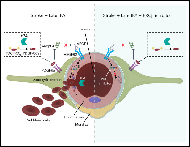
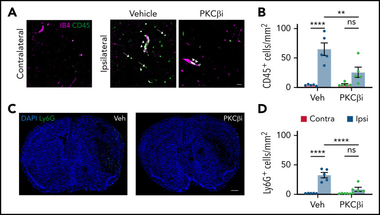
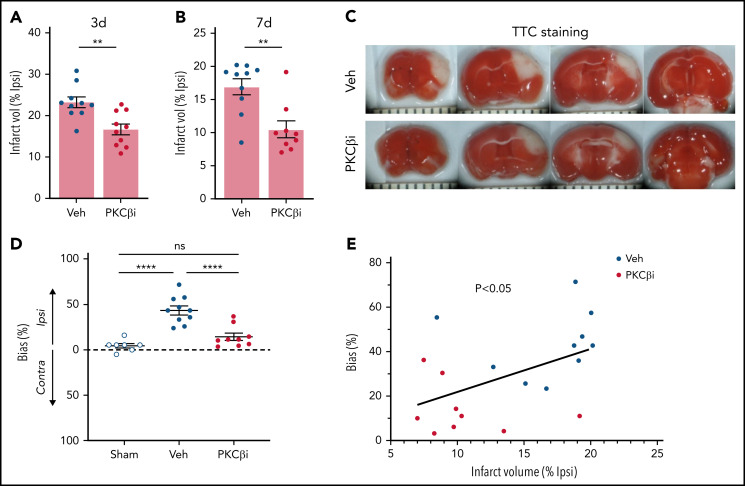
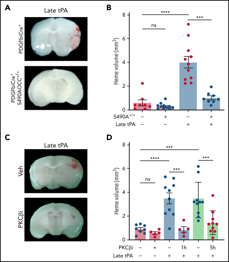
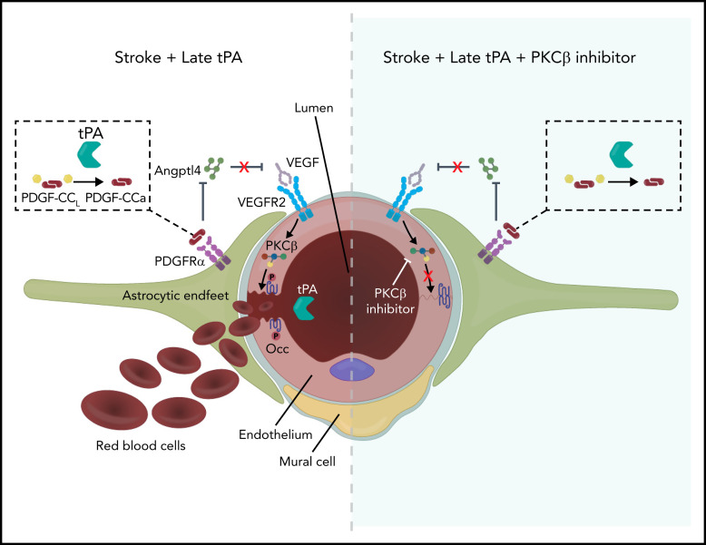
The current standard of care for moderate to severe ischemic stroke is thrombolytic therapy with tissue plasminogen activator (tPA). Treatment with tPA can significantly improve neurologic outcomes; however, thrombolytic therapy is associated with an increased risk of intracerebral hemorrhage (ICH). The risk of hemorrhage significantly limits the use of thrombolytic therapy, and identifying pathways induced by tPA that increase this risk could provide new therapeutic options to extend thrombolytic therapy to a wider patient population. Here, we investigate the role of protein kinase Cβ (PKCβ) phosphorylation of the tight junction protein occludin during ischemic stroke and its role in cerebrovascular permeability. We show that activation of this pathway by tPA is associated with an increased risk of ICH. Middle cerebral artery occlusion (MCAO) increased phosphorylation of occludin serine 490 (S490) in the ischemic penumbra in a tPA-dependent manner, as tPA-/- mice were significantly protected from MCAO-induced occludin phosphorylation. Intraventricular injection of tPA in the absence of ischemia was sufficient to induce occludin phosphorylation and vascular permeability in a PKCβ-dependent manner. Blocking occludin phosphorylation, either by targeted expression of a non-phosphorylatable form of occludin (S490A) or by pharmacologic inhibition of PKCβ, reduced MCAO-induced permeability and improved functional outcome. Furthermore, inhibiting PKCβ after MCAO prevented ICH associated with delayed thrombolysis. These results show that PKCβ phosphorylation of occludin is a downstream mediator of tPA-induced cerebrovascular permeability and suggest that PKCβ inhibitors could improve stroke outcome and prevent ICH associated with delayed thrombolysis, potentially extending the window for thrombolytic therapy in stroke.


Figures

None
Graphical abstract
Figure 1.
Figure 1.
MCAO induces occludin S490 phosphorylation in a tPA-dependent manner. Mice were subjected to MCAO and killed at 3 and 24 hours after MCAO. (A) Stitched images of the region adjacent to the ischemic core (Isch) showing vessel pS490 occludin (red) and total occludin (green) staining in WT mice compared with contralateral hemisphere. Scale bar, 60 µm. (B) Confocal images zoomed in on individual vessels showing pS490 occludin (red) and ZO-1 (white) staining in tPA−/− mice after MCAO compared with WT mice 24 hours’ post-MCAO. Scale bar, 17 µm. (C) Quantification of pS490 fluorescence intensity at the junction 24 hours after MCAO in WT and tPA−/− mice in the contralateral (Contra) and ipsilateral (Ipsi) regions. (D) The amount of pS490 and total occludin in extracts from the ipsilateral (I) and contralateral (C) hemispheres was determined by western blot in WT and tPA−/− mice at 3 and 24 hours’ post-MCAO. (E) Quantification is expressed as relative to WT contralateral from each time point. (F) ICV injection with either PBS or tPA was performed in WT mice; after 6 hours, protein levels of pS490 and total occludin were determined by western blot of the whole brain extract. (G) Quantification is shown. One-way analysis of variance followed by Holm-Šídák post hoc test was used for comparison of ≥3 groups; a t test was used for comparison between 2 groups. *P < .05, ***P < .001, ****P < .0001. ns, nonsignificant; Veh, vehicle.
Figure 2.
Figure 2.
tPA/PDGF-CCa signaling increases occludin phosphorylation and permeability. (A) Gene expression heatmap of the top differentially expressed genes in isolated vascular fragments from WT mice 4 hours after ICV injection of either PBS or PDGF-CCa. (B) The vascular fragments from WT mice were isolated 4 hours after ICV injection of PBS, PDGF-CCa, or tPA and were analyzed by using quantitative polymerase chain reaction for differential expression of Angptl4. (C) The isolated vascular fraction from WT mice, either control without MCAO or isolated 24 hours after MCAO, was analyzed by using quantitative polymerase chain reaction for the expression of Angptl4. (D) WT mice were given an ICV injection with PBS, tPA, or tPA in combination with imatinib, or ANGPTL4, or VEGFR2 inhibitor (VEGFR2i), or PKCβ inhibitor (PKCβi). At 5 hours, Evans blue was injected intravenously; at 6 hours, animals were perfused with PBS. One brain hemisphere was blotted to determine pS490 and total occludin protein levels. (E) Quantification of blot S490 phosphorylation. (F) The other brain hemisphere was processed for BBB permeability by measuring Evans blue dye extravasation. (G) BBB permeability was assessed after 6 hours of either saline or tPA ICV injection in PDGFiCre+ mice and PDGFiCre+; S490AOCC+/+ mice by measuring Evans blue dye extravasation. One-way analysis of variance followed by a Holm-Šídák post hoc test was used for comparison of ≥3 groups; a t test was used for comparison between 2 groups. *P < .05, **P < .01, ***P < .001, ****P < .0001. ns, nonsignificant; Veh, vehicle.
Figure 3.
Figure 3.
Inhibition of occludin S490 phosphorylation prevents MCAO-induced permeability. (A) PDGFiCre+ mice and PDGFiCre+; S490AOCC+/+ mice were subjected to MCAO, and BBB permeability was determined 24 hours later by quantifying a 70-kDa fluorescent dextran leak in cross-sections from both the ipsilateral (Ipsi) and the contralateral (Contra) region. (B) WT mice were given vehicle (Veh) or the PKCβ inhibitor (PKCβi; 10 mg/kg) once a day for 3 days and then subjected to MCAO followed by one additional dose of the inhibitor 1 hour later. pS490 occludin (red) and total occludin (green) were detected by immunostaining in brain sections 24 hours after MCAO. Scale bar, 25 µm. Quantification of pS490 at the junction (C) and total occludin from confocal images (D) 24 hours after MCAO. (E) Image of BBB permeability to 70-kDa dextran in entire coronal sections in both contralateral and ipsilateral hemispheres 24 hours after MCAO. Scale bar, 600 µm. (F) Quantification of dextran leak in cross-sections 24 hours after MCAO. (G) BBB permeability to 70-kDa dextran was also assessed by quantifying the amount of extravasated dye in brain homogenates 24 hours after MCAO. One-way analysis of variance followed by a Holm-Šídák post hoc test. *P < .05, ****P < .0001. ns, nonsignificant.
Figure 4.
Figure 4.
PKCβ inhibition reduces MCAO-induced inflammation. Mice were treated with either vehicle (Veh) or the PKCβ inhibitor (PKCβi; 10 mg/kg) once a day for 3 days and then subjected to MCAO followed by 1 additional dose of the inhibitor 1 hour later. (A) Twenty-four hours after MCAO, representative confocal images were obtained from the contralateral and ipsilateral (ischemic core) regions stained with vessel marker IB4 (purple) and leukocyte marker CD45 (green) showing leukostasis (arrowheads) and infiltration of CD45+ cells to the parenchyma (arrows). Scale bar, 30 µm. (C) Stitched confocal images of the whole brain section showing the contralateral region on the left and ipsilateral on the right, stained with neutrophil marker Ly6G (green) and nuclei marker 4′,6-diamidino-2-phenylindole (DAPI; blue). Scale bar, 500 µm. The number of CD45+ (B) and Ly6G+ (D) cells was counted in both contralateral (red bars) and ipsilateral (blue bars) hemispheres. Data are represented as mean ± standard error of the mean. One-way analysis of variance followed by a Holm-Šídák post hoc test. **P < .01, ****P < .0001. ns, nonsignificant.
Figure 5.
Figure 5.
PKCβ inhibition decreases infarct volume and improves functional outcome. WT mice were given vehicle or the PKCβ inhibitor (PKCβi; 10 mg/kg) once a day for 3 days and then subjected to MCAO followed by 3 additional daily doses of the inhibitor. Infarct volume was assessed by using 2,3,5-triphenyltetrazolium chloride (TTC) staining at 72 hours (A) and 7 days (B) after MCAO. (C) Representative images of TTC staining 7 days after MCAO. (D) Functional outcome was measured by assessing the lateralized bias in the corridor test 7 days after MCAO. (E) Pearson’s correlation between the lateralized bias and infarct volume in vehicle- and PKCβi-treated mice, 7 days after MCAO. One-way analysis of variance followed by a Holm-Šídák post hoc test was used for comparison of ≥3 groups; a t test was used for comparison between 2 groups. **P < .01, ****P < .0001. Contra, contralateral; Ipsi, ipsilateral; ns, nonsignificant; Veh, vehicle.
Figure 6.
Figure 6.
Inhibition of PKCβ prevents hemorrhagic transformation after delayed tPA treatment. (A) PDGFiCre+ mice and PDGFiCre+; S490AOCC+/+ mice were subjected to MCAO and then treated intravenously with either PBS or tPA (10 mg/kg) 5 hours after MCAO. (B) The volume of hemorrhage was quantified from serial brain sections 72 hours after MCAO. (C) Mice were subjected to MCAO; 1 hour (light blue bars) or 5 hours (green bars) later, animals were treated with vehicle (Veh) or PKCβ inhibitor (PKCβi; 10 mg/kg) daily for 3 days. Delayed tPA thrombolysis was performed 5 hours after MCAO, and brains were analyzed at 72 hours after MCAO. (D) Hemorrhage volume was measured 72 hours after MCAO. One-way analysis of variance followed by a Holm-Šídák post hoc test was used for comparison of ≥3 groups; a t test was used for comparison between 2 groups. ***P < .001, ****P < .0001. ns, nonsignificant.
Figure 7.
Figure 7.
Model of tPA signaling to occludin phosphorylation and BBB permeability. Ischemic stroke induces release of endogenous tPA that cleaves latent PDGF-CCL to active PDGF-CCa, which induces PDGFRα signaling in perivascular astrocytes. This results in decreased expression of ANGPTL4, relieving the ANGPTL4 inhibition of VEGF signaling, which further promotes ischemia-induced VEGF signaling through VEGFR2, which leads to activation of PKCβ and occludin phosphorylation on S490. This phosphorylation site regulates endocytosis of occludin with other junctional proteins, resulting in increased paracellular permeability. Recombinant thrombolytic tPA may leak into the brain parenchyma and further activate this system, leading to ICH when given beyond 4.5 hours after stroke. However, inhibiting PKCβ may provide a therapeutic option to maintain the BBB, extending the tPA therapeutic window. Note: Mural cell was retracted to emphasize VEGFR2 on the endothelial cell abluminal membrane.

Comment in

References

    1. Ahmad FB, Anderson RN. The leading causes of death in the US for 2020. JAMA. 2021;325(18):1829-1830. - PMC - PubMed
    1. Kochanek KD, Murphy SL, Xu J, Tejada-Vera B. Deaths: final data for 2014. Natl Vital Stat Rep. 2016;65(4):1-122. - PubMed
    1. Mozaffarian D, Benjamin EJ, Go AS, et al. ; Stroke Statistics Subcommittee . Heart Disease and Stroke Statistics–2016 update: a report from the American Heart Association. Circulation. 2016;133(4):e38-e360. - PubMed
    1. Keep RF, Hua Y, Xi G. Intracerebral haemorrhage: mechanisms of injury and therapeutic targets. Lancet Neurol. 2012; 11(8):720-731. - PMC - PubMed
    1. Hacke W, Donnan G, Fieschi C, et al. ; NINDS rt-PA Study Group Investigators . Association of outcome with early stroke treatment: pooled analysis of ATLANTIS, ECASS, and NINDS rt-PA stroke trials. Lancet. 2004; 363(9411):768-774. - PubMed

Publication types