. 2022 Jul:347:476-488.
doi: 10.1016/j.jconrel.2022.05.023. Epub 2022 May 20.

Nanoparticle-delivered TLR4 and RIG-I agonists enhance immune response to SARS-CoV-2 subunit vaccine

Affiliations

Nanoparticle-delivered TLR4 and RIG-I agonists enhance immune response to SARS-CoV-2 subunit vaccine

Alexandra Atalis et al. J Control Release. 2022 Jul.

Abstract

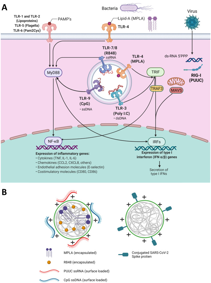
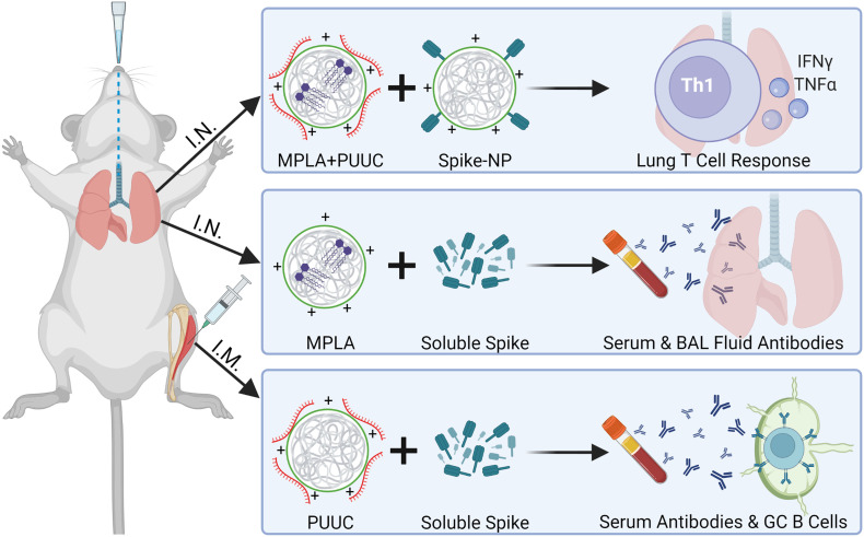
Despite success in vaccinating populations against SARS-CoV-2, concerns about immunity duration, continued efficacy against emerging variants, protection from infection and transmission, and worldwide vaccine availability remain. Molecular adjuvants targeting pattern recognition receptors (PRRs) on antigen-presenting cells (APCs) could improve and broaden the efficacy and durability of vaccine responses. Native SARS-CoV-2 infection stimulates various PRRs, including toll-like receptors (TLRs) and retinoic acid-inducible gene I (RIG-I)-like receptors. We hypothesized that targeting PRRs using molecular adjuvants on nanoparticles (NPs) along with a stabilized spike protein antigen could stimulate broad and efficient immune responses. Adjuvants targeting TLR4 (MPLA), TLR7/8 (R848), TLR9 (CpG), and RIG-I (PUUC) delivered on degradable polymer NPs were combined with the S1 subunit of spike protein and assessed in vitro with isogeneic mixed lymphocyte reactions (isoMLRs). For in vivo studies, the adjuvant-NPs were combined with stabilized spike protein or spike-conjugated NPs and assessed using a two-dose intranasal or intramuscular vaccination model in mice. Combination adjuvant-NPs simultaneously targeting TLR and RIG-I receptors (MPLA+PUUC, CpG+PUUC, and R848+PUUC) differentially induced T cell proliferation and increased proinflammatory cytokine secretion by APCs in vitro. When delivered intranasally, MPLA+PUUC NPs enhanced CD4+CD44+ activated memory T cell responses against spike protein in the lungs while MPLA NPs increased anti-spike IgA in the bronchoalveolar (BAL) fluid and IgG in the blood. Following intramuscular delivery, PUUC NPs induced strong humoral immune responses, characterized by increases in anti-spike IgG in the blood and germinal center B cell populations (GL7+ and BCL6+ B cells) in the draining lymph nodes (dLNs). MPLA+PUUC NPs further boosted spike protein-neutralizing antibody titers and T follicular helper cell populations in the dLNs. These results suggest that protein subunit vaccines with particle-delivered molecular adjuvants targeting TLR4 and RIG-I could lead to robust and unique route-specific adaptive immune responses against SARS-CoV-2.

Keywords: Adaptive immune response; COVID-19 protein subunit vaccine; Combination adjuvant; Intranasal versus intramuscular vaccination; Monophosphoryl lipid A; SARS-CoV-2 spike protein.


Conflict of interest statement

There are no conflicts of interest to disclose.

Figures

Unlabelled Image
Graphical abstract
Fig. 1
Fig. 1
TLR and RIG-I signaling pathways intersect and can be activated by nanoparticles with encapsulated and surface-loaded pathogen-associated molecular patterns. A) Schematic of signaling pathways initiated by TLRs 1–9, and RIG-I receptors including adaptor proteins MyD88, TRIF, TRAF3, and MAVS and transcription factors NF-κB and various IRFs which regulate the transcription of proinflammatory genes. B) Left: Depiction of poly(lactic-co-glycolic acid)-polyethyleneimine nanoparticles (PLGA-PEI NPs) with encapsulated hydrophobic molecules (R848 or MPLA) and surface-loaded nucleic acids (CpG or PUUC) for adjuvant delivery during vaccination. Right: PLGA-PEI NPs with surface-loaded SARS-CoV-2 spike protein for antigen delivery during vaccination.
Fig. 2
Fig. 2
GM-CSF and FLT3L BMDCs secrete different cytokine profiles and activate CD8+ T cells in response to S1 protein with combination adjuvants. A) t-SNE plots of GM-CSF and FLT3L BMDCs with labeled clusters of APC subsets. Macrophages/Mo-DCs are CD64+ F4/80, Conventional DCs are CD11c+ MHCII+ CD64lo F4/80lo, Monocytes are Ly6Chi CD11c, Neutrophils are Ly6Ghi Ly6C+, plasmacytoid DCs are B220+ Ly6C+ CD11c+ MHCIIlo. BG) Cytokine concentrations (pg/mL) of IL-1β, IL-27, IL-12p70, IFN-β, and IFN-λ3 in supernatants of BMDC culture after incubation with adjuvanted NPs for 24 h. HI) Percentage of live CD3+CD4+ T cells or CD3+CD8+ T cells proliferating in presence of GM-CSF BMDCs, gated on diminished CFSE signal (Fig. S1). “No Adjuvant” condition is blank NPs. J, K) Percentage of live CD3+CD4+ T cells or CD3+CD8+ T cells proliferating in presence of FLT3L BMDCs. *p < 0.05. **p < 0.01, ***p < 0.001, ****p < 0.0001 based on a One-Way ANOVA and Tukey Test for multiple comparison.
Fig. 3
Fig. 3
MPLA+PUUC NPs increase T cell responses in the lung when delivered intranasally with spike protein. On days 0 (1st dose) and 28 (2nd dose), female BALB/c mice were immunized I.N. with unformulated (i.e., soluble) or NP-conjugated spike protein (1 μg) and PLGA-PEI NPs (4 mg) loaded with MPLA (24 μg), PUUC (17 μg), and MPLA+PUUC (20 μg, 17 μg). Mice were euthanized and lungs were collected on day 35, one week after the 2nd dose. Lung cells were restimulated with spike peptide pools for 6 h and stained for analysis by flow cytometry. Percentages of cells expressing A) CD4+CD44+ out of CD45+ cells, B) IFNγ+ out of CD4+CD44+ cells, C) TNFα+ out of CD4+CD44+ cells, D) CD69+CD103+ (tissue resident memory T cells) out of CD45+ cells. BAL fluid from vaccinated mice using soluble spike antigen was assayed for anti-spike E) IgG and F) IgA with ELISA. G) Sera were assayed for anti-spike IgG with ELISA (error bars represent the SEM). p values are *p ≤ 0.05, **p ≤ 0.01, ***p ≤ 0.001 calculated using AD) One-way ANOVA with Tukey post-hoc test, E, F) Kruskal-Wallis with Dunn's post-hoc test for nonparametric data, or G) Two-way ANOVA with Tukey post-hoc test.
Fig. 4
Fig. 4
PUUC NPs delivered intramuscularly with spike protein enhance humoral responses. Female BALB/c mice were immunized I.M. into both tibialis anterior muscles at day 0 (1st dose) with soluble spike protein at doses of 80 ng, 200 ng, 1000 ng with or without adjuvant-NPs (4 mg) loaded with PUUC (+P, 20 ng PUUC dose). Peripheral blood was sampled on day 26. On day 28, mice received a 2nd dose of protein subunit vaccines. Mice received the same formulations, except for two groups that received 80 ng spike protein as a 1st dose received 1000 ng spike protein as a 2nd antigen dose (80/1000 and 80/1000 +P). Mice were euthanized on day 36 for to collect blood and popliteal LNs. A) Anti-spike IgG in post-1st dose sera at various dilutions measured by absorbance at 450 nm during ELISA assays and B) comparison of area under the curve (AUC). CD) Anti-spike IgG in post-2nd dose sera measured by absorbance at 450 nm and comparison of AUC. E) ACE-2 signal measured by absorbance at 450 nm in spike protein neutralization assay with post-2nd dose sera. Absorbance was normalized to a blank well in each row of a 384 well plate to correct for plate effects. Lower absorbance values indicate higher spike-neutralizing antibody levels in sera. Percentages of cells expressing F) Bcl6+ out of B220+ cells, G) GL7+ out of B220+ cells and H) CXCR5+ out of B220 cells from combined popliteal lymph nodes. B,D) Normality was assessed with the Kolmogorov-Smirnov test. Statistical significance was determined with the Kruskal-Wallis test and Dunn's post-hoc test for multiple comparisons. EH) Statistical significance calculated with One-Way ANOVA and Tukey post-hoc test. *p ≤ 0.05, **p ≤ 0.01, ***p ≤ 0.001, ****p ≤ 0.0001 for all graphs.
Fig. 5
Fig. 5
Key takeaways from intranasal and intramuscular two-dose vaccination models. Top: MPLA-PUUC NPs administered with spike-NPs intranasally induce local T cell responses, characterized by increases in IFNγ+ and TNFα+ CD4 T cells and tissue resident memory cells after lung cell restimulation with spike peptide pools. Middle: MPLA NPs administered with soluble spike protein intranasally induce systemic and localized humoral responses, characterized by increases in serum and BAL fluid antibodies. Bottom: PUUC NPs injected with soluble spike protein intramuscularly induce systemic humoral responses, characterized by increases in serum antibodies and germinal center B cells in the popliteal lymph nodes.

Update of

Similar articles

Cited by

References

    1. R&D Blue Print . 2021. COVID-19 Vaccine Tracker and Landscape.
    1. Awate S., Babiuk L.A.B., Mutwiri G. Mechanisms of action of adjuvants. Front. Immunol. 2013;4:114. - PMC - PubMed
    1. Reed S.G., Orr M.T., Fox C.B. Key roles of adjuvants in modern vaccines. Nat. Med. 2013;19:1597–1608. - PubMed
    1. Saravia J., Chapman N.M., Chi H. Helper T cell differentiation. Cell. Mol. Immunol. 2019;16:634–643. - PMC - PubMed
    1. Maddur M.S., Bayry J. B cells drive Th2 responses by instructing human dendritic cell maturation. Oncoimmunology. 2015;4 e1005508. - PMC - PubMed

Publication types