

Dapagliflozin protects neural and vascular dysfunction of the retina in diabetes
- PMID: 35577387
- PMCID: PMC9114950
- DOI: 10.1136/bmjdrc-2022-002801
Dapagliflozin protects neural and vascular dysfunction of the retina in diabetes
Abstract
Introduction: Dapagliflozin, a sodium-glucose transporter inhibitor, effectively reduces blood glucose and is indicated for individuals with kidney diseases and cardiovascular disorders. In this study, we further expand the therapeutic benefit of dapagliflozin in the neural and vascular retina, with the potential to effectively manage diabetic retinopathy (DR), the most common complication of diabetes.
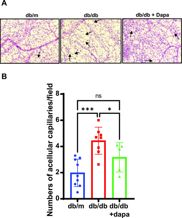
Research design and methods: Db/db mice, an animal model of type 2 diabetes, were treated with dapagliflozin orally, and the electroretinogram (ERG) response and acellular capillary numbers were assessed. Messenger RNA levels of inflammatory cytokines were studied using real-time quantitative (q)PCR. We assessed endothelial cell migration in a scratch wound assay and retinal glucose uptake using human retinal endothelial cells.
Results: The dapagliflozin treatment improved the ERG b-wave amplitude and decreased acellular capillary numbers. The scratch wound assay demonstrated a reduction in wound closure after dapagliflozin treatment. Retinal glucose uptake reduced after dapagliflozin treatment compared with the respective controls.
Conclusions: Our studies suggest that dapagliflozin treatment effectively corrects neural and vascular dysfunction of the retina in diabetes. This effect is mediated by a decrease in inflammation and improved glycemic control. In addition, dapagliflozin exhibits decreased wound healing and glucose uptake, which could benefit the retina. Thus, dapagliflozin could be helpful in the management of DR, with multimodal therapeutic effects.
Keywords: Diabetes Complications; Diabetes Mellitus, Type 2; Diabetic Retinopathy; Retina.
© Author(s) (or their employer(s)) 2022. Re-use permitted under CC BY-NC. No commercial re-use. See rights and permissions. Published by BMJ.
Conflict of interest statement
Competing interests: ADB is an ad hoc staff pharmacist at CVS Health/Aetna. The content of this study does not reflect those of CVS Health/Aetna. QL, SPL, EB, HD, and DM do not have any conflicts of interest with the study.
Figures


Similar articles
-
SGLT2 inhibition via dapagliflozin improves generalized vascular dysfunction and alters the gut microbiota in type 2 diabetic mice.Cardiovasc Diabetol. 2018 Apr 27;17(1):62. doi: 10.1186/s12933-018-0708-x. Cardiovasc Diabetol. 2018. PMID: 29703207 Free PMC article.
-
Dapagliflozin ameliorated retinal vascular permeability in diabetic retinopathy rats by suppressing inflammatory factors.J Diabetes Complications. 2024 Mar;38(3):108631. doi: 10.1016/j.jdiacomp.2023.108631. Epub 2023 Oct 17. J Diabetes Complications. 2024. PMID: 38340519
-
The Effect of Sodium-Dependent Glucose Cotransporter 2 Inhibitor Tofogliflozin on Neurovascular Coupling in the Retina in Type 2 Diabetic Mice.Int J Mol Sci. 2022 Jan 25;23(3):1362. doi: 10.3390/ijms23031362. Int J Mol Sci. 2022. PMID: 35163285 Free PMC article.
-
[Dapagliflozin, the first SGLT-2 inhibitor in the treatment of type 2 diabetes].Med Clin (Barc). 2013 Sep;141 Suppl 2:36-43. doi: 10.1016/S0025-7753(13)70062-9. Med Clin (Barc). 2013. PMID: 24444523 Review. Spanish.
-
Development of the sodium-glucose co-transporter 2 inhibitor dapagliflozin for the treatment of patients with type 2 diabetes mellitus.Expert Rev Clin Pharmacol. 2011 Nov;4(6):669-83. doi: 10.1586/ecp.11.54. Expert Rev Clin Pharmacol. 2011. PMID: 22111852 Review.
Cited by
-
BMAL1 Overexpression in Suprachiasmatic Nucleus Protects from Retinal Neurovascular Deficits in Diabetes.bioRxiv [Preprint]. 2025 Feb 6:2025.02.05.636648. doi: 10.1101/2025.02.05.636648. bioRxiv. 2025. PMID: 39975095 Free PMC article. Preprint.
-
Novel Antidiabetic Drugs and the Risk of Diabetic Retinopathy: A Systematic Review and Meta-Analysis of Randomized Controlled Trials.J Clin Med. 2024 Mar 20;13(6):1797. doi: 10.3390/jcm13061797. J Clin Med. 2024. PMID: 38542021 Free PMC article. Review.
-
The Impact of SGLT2 Inhibitors in the Heart and Kidneys Regardless of Diabetes Status.Int J Mol Sci. 2023 Sep 18;24(18):14243. doi: 10.3390/ijms241814243. Int J Mol Sci. 2023. PMID: 37762542 Free PMC article. Review.
References
Publication types
MeSH terms
Substances
Grants and funding
LinkOut - more resources
Full Text Sources
Medical
Miscellaneous