. 2022 Jun 8;42(23):4725-4736.
doi: 10.1523/JNEUROSCI.2273-21.2022. Epub 2022 May 16.

Voluntary Exercise Boosts Striatal Dopamine Release: Evidence for the Necessary and Sufficient Role of BDNF

Affiliations

Voluntary Exercise Boosts Striatal Dopamine Release: Evidence for the Necessary and Sufficient Role of BDNF

Guendalina Bastioli et al. J Neurosci. .

Abstract

Physical exercise improves motor performance in individuals with Parkinson's disease and elevates mood in those with depression. Although underlying factors have not been identified, clues arise from previous studies showing a link between cognitive benefits of exercise and increases in brain-derived neurotrophic factor (BDNF). Here, we investigated the influence of voluntary wheel-running exercise on BDNF levels in the striatum of young male wild-type (WT) mice, and on the striatal release of a key motor-system transmitter, dopamine (DA). Mice were allowed unlimited access to a freely rotating wheel (runners) or a locked wheel (controls) for 30 d. Electrically evoked DA release was quantified in ex vivo corticostriatal slices from these animals using fast-scan cyclic voltammetry. We found that exercise increased BDNF levels in dorsal striatum (dStr) and increased DA release in dStr and in nucleus accumbens core and shell. Increased DA release was independent of striatal acetylcholine (ACh), and persisted after a week of rest. We tested a role for BDNF in the influence of exercise on DA release using mice that were heterozygous for BDNF deletion (BDNF+/-). In contrast to WT mice, evoked DA release did not differ between BDNF+/- runners and controls. Complementary pharmacological studies using a tropomyosin receptor kinase B (TrkB) agonist in WT mouse slices showed that TrkB receptor activation also increased evoked DA release throughout striatum in an ACh-independent manner. Together, these data support a causal role for BDNF in exercise-enhanced striatal DA release and provide mechanistic insight into the beneficial effects of exercise in neuropsychiatric disorders, including Parkinson's, depression, and anxiety.SIGNIFICANCE STATEMENT Exercise has been shown to improve movement and cognition in humans and rodents. Here, we report that voluntary exercise for 30 d leads to an increase in evoked DA release throughout the striatum and an increase in BDNF in the dorsal (motor) striatum. The increase in DA release appears to require BDNF, indicated by the absence of DA release enhancement with running in BDNF+/- mice. Activation of BDNF receptors using a pharmacological agonist was also shown to boost DA release. Together, these data support a necessary and sufficient role for BDNF in exercise-enhanced DA release and provide mechanistic insight into the reported benefits of exercise in individuals with dopamine-linked neuropsychiatric disorders, including Parkinson's disease and depression.

Keywords: Parkinson's disease; brain slices; fast-scan cyclic voltammetry; nAChRs; nucleus accumbens; running wheel.


Figures

Figure 1.
Figure 1.
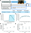
Voluntary wheel running. A, Timeline for voluntary wheel-running protocol; for each study, 12 randomly assigned mice were housed individually with either a freely rotating wheel (runners; n = 6) or a locked wheel (controls; n = 6). After each 30 d study, brain tissue was collected for HPLC analysis of DA and DOPAC tissue content, Western blotting (WB) for BDNF expression, or FSCV for evaluation of evoked DA release. B, Average time course of wheel running activity for three cohorts of 6 mice each (n = 18 runners) showing diurnal variation, with greater activity in the dark phase (shaded light blue) than in the light. C, Average total running per day (n = 18 runners). D, Change in body weight monitored weekly, with the first day of housing with a wheel taken as baseline 100%, shows a comparable increase in weight over the experimental period between runners and controls (p < 0.0001 initial vs. final weight, n = 18 mice per group, unpaired t test). E, Average weekly food consumption for three cohorts assessed weekly (*p < 0.05, **p < 0.01, ***p < 0.001 runners vs controls; n = 18 per group in study; 2-way ANOVA, Bonferroni post hoc test).
Figure 2.
Figure 2.
DA and DOPAC tissue contents and BDNF expression in dStr and vStr after 30 d of voluntary wheel running. A, B, Tissue content of DA in dStr and vStr did not differ between runners and controls (n = 26 samples from 6 mice per group; unpaired t test). C, D, Tissue content of the DA metabolite DOPAC did not differ between runners and controls in either dStr or vStr (n = 24–29 samples from 6 mice per group; unpaired t test). E, F, BDNF expression in dStr and vStr after 30 d wheel running; quantitative data were normalized to β-actin (*p < 0.05 runners vs controls; n = 6 mice per group, 1 sample per mouse; unpaired t test).
Figure 3.
Figure 3.
Increased evoked [DA]o in dStr, NAc core and NAc shell in ex vivo striatal slices after 30 d of voluntary wheel running. A, Left, Coronal section of mouse brain showing typical level of forebrain slices used to study axonal DA release (modified from Franklin and Paxinos, 2008). At this level, local electrical stimulation can be used to evoke DA release in dorsolateral dStr and in the NAc core and shell in the same slice. Right, Representative voltammogram recorded in the dStr following local, single-pulse stimulation (1 pulse), showing characteristic DA oxidation (+0.61 vs AgAgCl) and reduction (−0.24 V vs Ag/AgCl) peak potentials; similar voltammograms were obtained in NAc core and shell. BD, Left, Average evoked increases in [DA]o in dStr, NAc core (single-pulse stimulation) and NAc shell (5 pulse, 100 Hz) in ex vivo slices from runners and controls, normalized to mean peak [DA]o for each region in controls (error bars omitted); arrow indicates time of stimulation. Right, Data summary for evoked [DA]o in each region for runners versus controls (n = 60–103 sites per region, 2 slices per mouse, 12 mice per group; unpaired U or t tests). E, F, Left, Average evoked increases in [DA]o in dStr and NAc core (single-pulse stimulation) in the same slices examined in B–D after superfusion of DHβE (1 μm), a nAChR antagonist. Data are normalized to mean peak evoked [DA]o in DHβE for each region in controls. Right, Data summary; evoked [DA]o, dStr, and NAc core in the presence of DHβE (n = 29–42 sites per region, 2 slices per mouse, 6 mice per group; unpaired U tests). B–F, *p < 0.05, **p < 0.01, ***p < 0.001.
Figure 4.
Figure 4.
Enduring enhancement of evoked [DA]o in dStr, NAc core, and NAc shell after 7 d rest. A, Timeline for voluntary wheel running for 30 d followed by 7 d of rest (locked running wheel for runners as well as controls; n = 4 mice per group). B–D, Left, Average evoked increases in [DA]o in dStr, NAc core, and NAc shell in slices from runners and controls, normalized to mean peak [DA]o for each region in controls (error bars omitted). Right, Data summary; evoked [DA]o remained higher in runners than controls in dStr and NAc core, but the difference in NAc shell was lost (n = 40–80 sites per region, 4 slices per mouse, 4 mice per group; unpaired U tests). E, F, Left, Averaged evoked increases in [DA]o in dStr and NAc in the presence of DHβE (1 μm), normalized to mean peak evoked [DA]o in DHβE for each region in controls. Right, Data summary; evoked [DA]o in DHβE in dStr and NAc core from slices for runners versus controls (n = 68–80 sites per region, 4 slices per mouse, 4 mice per group; unpaired, one-tailed U tests). B–F, *p < 0.05, **p < 0.01, ***p < 0.001.
Figure 5.
Figure 5.
Voluntary wheel running in BDNF+/− mice. A, Average time course of running-wheel activity for WT mice (Figure 1B, blue line) and BDNF+/− mice (green line) shows diurnal variation, with greater activity in the dark phase (shaded light blue) than in the light, albeit with a different pattern than seen in WT mice (***p < 0.001; n = 18 WT mice, n = 6 BDNF+/− mice; 2-way ANOVA, Bonferroni post hoc test). B, Average total daily running over the 30 d running period did not differ between WT mice (Figure 1C) and BDNF+/− mice (2-way ANOVA, Bonferroni post hoc test; n = 18 WT mice, n = 6 BDNF+/− mice). C, Body weight of BDNF+/− mice did not change over the running period for either runner or control BDNF+/− mice and did not differ between runners and controls at any point during the running period; the first day of the running period was taken as baseline (runners vs controls; n = 6 mice per group; 2-way ANOVA, Bonferroni post hoc test). D, Average weekly food consumption also did not differ between runner and control BDNF+/− mice (runners vs controls; n = 6 mice per group; 2-way ANOVA, Bonferroni post hoc test). E, F, BDNF expression in dStr and vStr on the last day of the 30 d wheel running did not differ between runner and control BDNF+/− mice; quantitative data normalized to β-actin (n = 6 mice per group, runners vs controls; unpaired t test); dashed lines indicate average BDNF content for the corresponding striatal region from WT mice.
Figure 6.
Figure 6.
Loss of effect of voluntary wheel running on nigrostriatal DA release in BDNF+/− mice. A–C, Left, Average evoked [DA]o in dStr, NAc core, and NAc shell, normalized to mean peak evoked [DA]o for each region in controls. Right, Data summary for BDNF+/− runners vs controls (n = 40–57 sites, 2 slices per mouse, 6 mice per group; unpaired t test). D, E, Left, Average evoked [DA]o in dStr and NAc core (error bars omitted) in the same slices examined in A–C after superfusion of DHβE (1 μm). Right, Data summary for evoked increases in [DA]o in the presence of DHβE in slices from BDNF+/− runners and controls (n = 37–49 sites, 2 slices per mouse, 6 mice per group; unpaired t or U tests). C, E, *p < 0.05, **p < 0.01.
Figure 7.
Figure 7.
Activation of TrkB receptors in ex vivo striatal slices enhances evoked [DA]o. A–D, Summary of peak evoked [DA]o in dStr, in ex vivo slices after 2 h exposure to LM22-A4 (1 μm; a TrkB receptor agonist), to LM22-4A + DHβE (1 μm), to LM22-4A + LY29004 (1 μm), to LM22-4A + U73122 (1 μm), and in time-matched control slices with aCSF alone or the corresponding inhibitor, normalized to mean peak [DA]o in each region for time-matched controls (n = 24–30 sites per region, 2 slices per mouse, 3 mice per group; unpaired t tests). E–H, Summary of peak evoked [DA]o in NAc core; in ex vivo slices after 2 h exposure to LM22-A4, to LM22-4A + DHβE, to LM22-4A + LY29004, and to LM22-4A + U73122; and in time-matched control slices, every group with corresponding inhibitor, normalized to mean peak [DA]o in each region for controls (n = 27–37 sites, 2 slices per mouse, 3 mice per group; unpaired t tests). I–K, Summary of peak evoked [DA]o in NAc shell; in ex vivo slices after 2 h exposure to LM22-A4, to LM22-4A + LY29004, and to LM22-4A + U73122; and in time-matched control slices, each group with the corresponding inhibitor, normalized to mean peak [DA]o in each region for controls (unpaired t test, n = 18–30 sites, 2 slices per mouse, 3 mice per group; unpaired t tests). A–G, *p < 0.05, **p < 0.01, ***p < 0.001.

References

    1. Aguiar AS Jr, Tristão FS, Amar M, Chevarin C, Glaser V, de Paula Martins R, Moreira EL, Mongeau R, Lanfumey L, Raisman-Vozari R, Latini A, Prediger RD (2014) Six weeks of voluntary exercise don't protect C57BL/6 mice against neurotoxicity of MPTP and MPP+. Neurotox Res 25:147–152. 10.1007/s12640-013-9412-5 - DOI - PubMed
    1. Ahlskog JE (2018) Aerobic exercise: evidence for a direct brain effect to slow Parkinson disease progression. Mayo Clin Proc 93:360–372. 10.1016/j.mayocp.2017.12.015 - DOI - PubMed
    1. Albeck DS, Sano K, Prewitt GE, Dalton L (2006) Mild forced treadmill exercise enhances spatial learning in the aged rat. Behav Brain Res 168:345–348. 10.1016/j.bbr.2005.11.008 - DOI - PubMed
    1. Albin RL, Young AB, Penney JB (1989) The functional anatomy of basal ganglia disorders. Trends Neurosci 12:366–375. 10.1016/0166-2236(89)90074-x - DOI - PubMed
    1. Altar CA, Cai N, Bliven T, Juhasz M, Conner JM, Acheson AL, Lindsay RM, Wiegand SJ (1997) Anterograde transport of brain-derived neurotrophic factor and its role in the brain. Nature 389:856–860. 10.1038/39885 - DOI - PubMed

Publication types