. 2022 Jun;33(6):1077-1086.
doi: 10.1681/ASN.2021121548. Epub 2022 May 16.

Arginase-1 Is Required for Macrophage-Mediated Renal Tubule Regeneration

Affiliations

Arginase-1 Is Required for Macrophage-Mediated Renal Tubule Regeneration

Naomi S Shin et al. J Am Soc Nephrol. 2022 Jun.

Abstract

Background: After kidney injury, macrophages transition from initial proinflammatory activation to a proreparative phenotype characterized by expression of arginase-1 (Arg1), mannose receptor 1 (Mrc1), and macrophage scavenger receptor 1 (Msr1). The mechanism by which these alternatively activated macrophages promote repair is unknown.

Methods: We characterized the macrophage and renal responses after ischemia-reperfusion injury with contralateral nephrectomy in LysM-Cre;Arg1fl/fl mice and littermate controls and used in vitro coculture of macrophages and tubular cells to determine how macrophage-expressed arginase-1 promotes kidney repair.

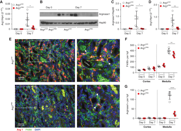
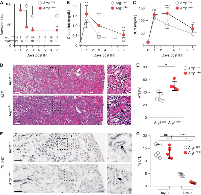
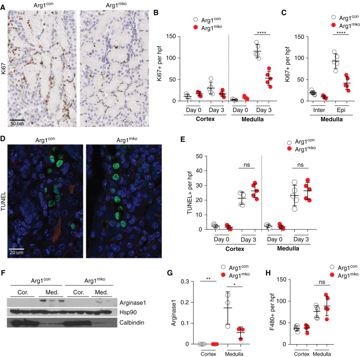
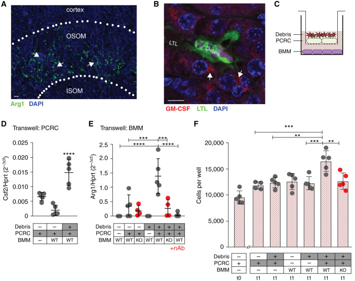
Results: After ischemia-reperfusion injury with contralateral nephrectomy, Arg1-expressing macrophages were almost exclusively located in the outer stripe of the medulla adjacent to injured S3 tubule segments containing luminal debris or casts. Macrophage Arg1 expression was reduced by more than 90% in injured LysM-Cre;Arg1fl/fl mice, resulting in decreased mouse survival, decreased renal tubular cell proliferation and decreased renal repair compared with littermate controls. In vitro studies demonstrate that tubular cells exposed apically to dead cell debris secrete high levels of GM-CSF and induce reparative macrophage activation, with those macrophages in turn secreting Arg1-dependent factor(s) that directly stimulate tubular cell proliferation.

Conclusions: GM-CSF-induced, proreparative macrophages express arginase-1, which is required for the S3 tubular cell proliferative response that promotes renal repair after ischemia-reperfusion injury.

Keywords: arginase-1; kidney repair; kidney tubules; macrophage; regeneration.


Figures

Figure 1.
Figure 1.
Arg1fl/fl;LysMCre/+ mice have reduced arginase-1 in the kidney after ischemic injury. Eight-week-old male Arg1mko (Arg1fl/fl;LysMCre/+) mice underwent IRI, and kidney lysates were collected for RNA and protein on day 0 and day 7 after surgery. (A) Arg1 mRNA expression level (relative to Hprt1) was significantly decreased in Arg1mko mice at day 7 within the whole kidney (*P<0.05, t test). (B and C) Western blot analysis (B) demonstrates significantly decreased arginase-1 protein levels on day 7 after IRI in Arg1mko mice, quantified in (C). Each lane represents one mouse kidney (*P<0.05, t test). (D) CD45+F4/80+ macrophages were isolated from kidneys at the indicated time by FACS and Arg1 mRNA expression quantified by quantitative rtPCR. Arg1 mRNA is significantly lower in day 7 macrophages from Arg1mko compared with control mice (**P<0.01, t test). (E) Immunofluorescence staining of injured kidneys from Arg1con mice (top panels) and Arg1mko mice (bottom panels) on day 7 after IRI (arginase-1 [red], F4/80+ [green], DAPI [blue]) shows significantly reduced arginase-1 staining in F4/80+ macrophages from the Arg1mko mouse. The boxed area in the left hand panels is magnified in the right panels. Asterisks indicate arginase-1+ and F4/80+ macrophage; arrows indicate arginase-1 and F4/80+ macrophage. (F) Quantification of total F4/80+ cells demonstrates increased medullary macrophages on day 7 after IRI Arg1con mice with a modest reduction seen in Arg1mko mice. (**P<0.01, ANOVA). (G) Quantification of arginase-1+ cells shows an 84% reduction in the Arg1mko mice. (****P<0.0001, ANOVA). n=5 mice per group. Casts exhibit green autofluorescence.
Figure 2.
Figure 2.
Macrophage expression of Arg1 is required for renal recovery. Arg1con (n=21) and Arg1mko mice (n=27) were subjected to 27 minutes of IRI/CL-NX. (A) Survival was significantly decreased in Arg1mko mice compared with control mice (**P<0.01, Log-rank Mantel–Haenszel test). (B) Serum creatinine and (C) BUN are equally increased on day 1, but reflect reduced recovery of GFR in the surviving Arg1mko mice on day 3 (****P<0.0001, ***P<0.001, ANOVA). (D) Histology demonstrates persistent injury in the Arg1mko mice on day 7 after IRI compared with control. Boxed area in the left panel is magnified in the right panel (gray arrow indicates clear tubule; black arrow indicates cast-filled tubule). (E) Acute tubular injury (ATI) score of kidneys from five mice on day 7 after IRI (**P<0.01, t test). (F) LTL staining of brush border at 7 days post IRI. (G) Quantification of LTL staining on days 0 and 7 shows reduced recovery of proximal tubule brush border in Arg1mko mice (****P<0.0001, *P<0.05, ANOVA). Scale bar, 100 μm.
Figure 3.
Figure 3.
Arginase-1+ macrophages promote epithelial cell proliferation. (A–C) Ki67 staining of Arg1con and Arg1mko kidneys on day 3 after IRI/CL-NX is shown in (A) and quantified in (B) (total cells) and (C) (interstitial and epithelial cells). Outer medullary Ki67+-proliferating epithelial cells are significantly reduced in Arg1mko kidneys (****P<0.0001, ANOVA). (D and E) TUNEL staining of Arg1con and Arg1mko kidneys on day 3 after IRI/CL-NX is shown in (D) and quantified in (E) (ns, not significant; t test). (F and G) Western blot analysis of the cortex and medulla of Arg1con and Arg1mko kidneys 3 days after IRI/CL-NX reveals upregulation of arginase-1 selectively in the medulla (F), with a significant reduction in the Arg1mko kidneys (G) (**P<0.01, *P<0.05, ANOVA). (H) Quantification of F4/80+ cells 3 days after IRI/CL-NX demonstrates no difference in macrophage numbers in the two groups. n=5 mice per group.
Figure 4.
Figure 4.
Luminal debris induces bidirectional tubular cell:macrophage cross-talk to promote tubule proliferative repair. (A) Immunofluorescent staining of mouse kidney for F4/80 after IRI/CL-NX reveals that arginase-1 (green) is limited to the outer stripe of the outer medulla near sites of intraluminal casts. (OSOM, outer stripe of the outer medulla; ISOM, inner stripe of the outer medulla; arrows indicate casts). Scale bar, 20 μm. (B) Immunofluorescent staining of mouse kidney for GM-CSF shows expression in epithelial cells adjacent to intraluminal cell debris (casts) after IRI/CL-NX (arrows indicate GM-CSF staining, LTL indicates brush border staining). Scale bar, 10 μm. (C) Schematic of Transwell experiments showing PCRCs and cultured naïve BMMs. (D) Csf2 mRNA expression is significantly higher in PCRCs cultured with cell debris on the apical surface (****P<0.0001, ANOVA). (E) Arg1 mRNA expression is significantly higher in wild-type (WT) BMMs cocultured with PCRCs exposed to debris versus PCRCs alone. Arg1 expression is significantly reduced in Arg1mko macrophages (KO) (****P<0.0001, ***P<0.001, ANOVA). (F) PCRCs were seeded at 10,000 cells per well (t0) and treated with conditioned media from Transwell cultures containing the components indicated under each bar, and cell numbers were counted at 24 hours (t1) (***P<0.001, **P<0.01, ANOVA).

Comment in

References

    1. Day YJ, Huang L, Ye H, Linden J, Okusa MD: Renal ischemia-reperfusion injury and adenosine 2A receptor-mediated tissue protection: Role of macrophages. Am J Physiol Renal Physiol 288: F722–F731, 2005 - PubMed
    1. Li L, Okusa MD: Macrophages, dendritic cells, and kidney ischemia-reperfusion injury. Semin Nephrol 30: 268–277, 2010 - PMC - PubMed
    1. Lee S, Huen S, Nishio H, Nishio S, Lee HK, Choi BS, et al. : Distinct macrophage phenotypes contribute to kidney injury and repair. J Am Soc Nephrol 22: 317–326, 2011 - PMC - PubMed
    1. Jo SK, Sung SA, Cho WY, Go KJ, Kim HK: Macrophages contribute to the initiation of ischaemic acute renal failure in rats. Nephrol Dial Transplant 21: 1231–1239, 2006 - PubMed
    1. Huen SC, Huynh L, Marlier A, Lee Y, Moeckel GW, Cantley LG: GM-CSF promotes macrophage alternative activation after renal ischemia/reperfusion injury. J Am Soc Nephrol 26: 1334–1345, 2015 - PMC - PubMed

Publication types

Substances