. 2022 May 16;12(1):8044.
doi: 10.1038/s41598-022-11601-1.

Evaluation of anterior cruciate ligament surgical reconstruction through finite element analysis

Affiliations

Evaluation of anterior cruciate ligament surgical reconstruction through finite element analysis

Konstantinos Risvas et al. Sci Rep. .

Abstract

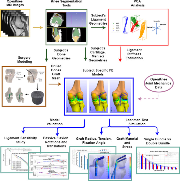
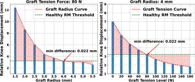
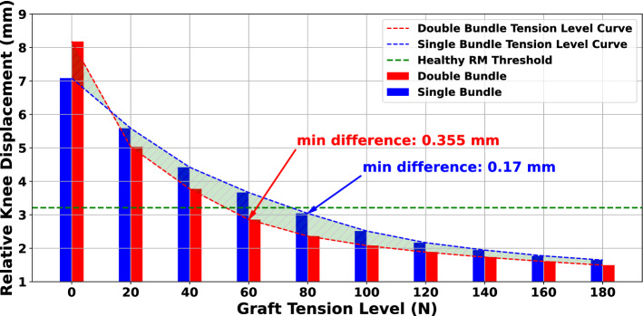
Anterior cruciate ligament (ACL) tear is one of the most common knee injuries. The ACL reconstruction surgery aims to restore healthy knee function by replacing the injured ligament with a graft. Proper selection of the optimal surgery parameters is a complex task. To this end, we developed an automated modeling framework that accepts subject-specific geometries and produces finite element knee models incorporating different surgical techniques. Initially, we developed a reference model of the intact knee, validated with data provided by the Open Knee(s) project. This helped us evaluate the effectiveness of estimating ligament stiffness directly from MRI. Next, we performed a plethora of "what-if" simulations, comparing responses with the reference model. We found that (a) increasing graft pretension and radius reduces relative knee displacement, (b) the correlation of graft radius and tension should not be neglected, (c) graft fixation angle of 20[Formula: see text] can reduce knee laxity, and (d) single-versus double-bundle techniques demonstrate comparable performance in restraining knee translation. In most cases, these findings confirm reported values from comparative clinical studies. The numerical models are made publicly available, allowing for experimental reuse and lowering the barriers for meta-studies. The modeling approach proposed here can complement orthopedic surgeons in their decision-making.


Conflict of interest statement

The authors declare no competing interests.

Figures

Figure 1
Figure 1
Overview of the proposed workflow. MRI data are used to propose a surgery plan for subject-specific ACLR. First, we use open-source segmentation tools to acquire geometries of the anatomical structures. Next, PCA is applied to ligament geometries to acquire subject-specific estimation of stiffness (PCA block). The bone geometries are used in the Surgery Modeling tool to drill the tunnels. During this process, the graft mesh is also generated. Subsequently, we automatically generate FE models of the knee joint. Joint mechanics data from the open-source Open Knee(s) project are used to develop and validate a RM. Then, the surgery parameters are evaluated by performing a FE simulation of the Lachman clinical examination. The performance of models that correspond to the ACL reconstructed knee joint are compared to that of the RM. The measurements of interest are relative knee displacement and graft maximum principal stress (Diagrams.net, v17.2.1, https://www.diagrams.net/).
Figure 2
Figure 2
Overview of ACLR surgery modeling workflow. The bone tunnels are “drilled” using cylindrical objects that trace a NURBS curve. (a) The NURBS curve is defined based on the anatomical landmarks, (b) cylindrical meshes are used for the tunnel “drilling” procedure, (c) the “drilled” tibia and femoral tunnels, and (d) the graft is morphed to the path and placed precisely through the anatomical landmarks.
Figure 3
Figure 3
FE model versions. In this work, we developed three different versions of a FE knee model that correspond to (a) the healthy knee used for reference and comparison (RM), (b) the ACL reconstructed knee with the SB approach, and (c) the DB technique.
Figure 4
Figure 4
Estimating ligament stiffness from MRI. Cross-sections and the bounding box of each ligament surface mesh are presented. Multiple cross-sections are taken along the principal ligament axis derived by PCA. The ligament’s cross-sectional area is estimated as the mean of the areas of each slice.
Figure 5
Figure 5
Aggregated results from the sensitivity analysis with the ten best combinations of stiffness and prestrain values for ACL and PCL. The estimated stiffness (PCA method) and recommended prestrain value from Blakenvoort were used as initial guesses. The “s” and “p” denote the stiffness and prestrain percentage change, respectively. In addition, we observed combinations that include the initial stiffness value and changed only the prestrain by a factor.
Figure 6
Figure 6
Comparison of simulated and experimental kinematics during passive knee flexion (translations left and rotations right). We observe that the proposed FE model exhibits comparable performance for the translations except for the medial-lateral direction. Regarding rotations, different behavior is evident for the internal-external rotation with a good initial match during varus-valgus rotation.
Figure 7
Figure 7
Effect of graft radius and pretension on knee laxity for a semitendinosus graft. The “difference” term refers to the absolute difference in relative knee displacement between each ACLR FE model and the RM. Increasing the graft radius for a specific pretension load reduces relative displacement. The same applies when increasing graft pretension for a fixed value of graft radius.
Figure 8
Figure 8
Comparison of three graft materials for different pretension and graft radius values. The absolute difference in relative knee displacement between each ACLR FE model and the RM was annotated using contour lines. Combinations of graft tension and graft radius that are close to the RM are inside the area with an absolute difference below 0.5, highlighted with dark blue color.
Figure 9
Figure 9
Effect of graft fixation angle on relative knee displacement. The “difference” term refers to the absolute difference in relative knee displacement between the ACLR FE model and the RM. It is noticed that for angles larger than 30 the relative knee laxity increases. In our case, we found the optimal fixation angles are between 15 and 20.
Figure 10
Figure 10
Results for comparing the SB and DB techniques. The “difference” term refers to the absolute difference in relative knee displacement translation between the FE model and the RM. Although the DB method appears to be superior in restraining knee laxity, the margin between the two methods is not disproportionate.

Similar articles

Cited by

References

    1. Laskowski, E. Acl injury and rehabilitation. Curr. Phys. Med. Rehabil. Rep.2. 10.1007/s40141-013-0036-8 (2014).
    1. Wetters, N., Weber, A., Wuerz, T., Schub, D. & Mandelbaum, B. Mechanism of injury and risk factors for anterior cruciate ligament injury. Oper. Tech. Sp. Med.24. 10.1053/j.otsm.2015.09.001 (2015).
    1. II, J., Miller, L. & Block, J. Quality of life in patients with knee osteoarthritis: A commentary on nonsurgical and surgical treatments. Open Orthopaed. J.7, 619–623. 10.2174/1874325001307010619 (2013). - PMC - PubMed
    1. Mahapatra P, Horriat S, Anand B. Anterior cruciate ligament repair -past, present and future. J. Exp. Orthopaed. 2018;5:20. doi: 10.1186/s40634-018-0136-6. - DOI - PMC - PubMed
    1. Legnani C, Ventura A, Terzaghi C, Borgo E, Albisetti W. Anterior cruciate ligament reconstruction with synthetic grafts. A review of literature. Int. Orthop. 2010;34:465–71. doi: 10.1007/s00264-010-0963-2. - DOI - PMC - PubMed

Publication types

MeSH terms