

doi: 10.1038/s41598-022-11830-4.
A high resolution scanning electron microscopy analysis of intracranial thrombi embedded along the stent retrievers
Affiliations
- PMID: 35577906
- PMCID: PMC9110407
- DOI: 10.1038/s41598-022-11830-4
Item in Clipboard
A high resolution scanning electron microscopy analysis of intracranial thrombi embedded along the stent retrievers
Sci Rep.
.
Abstract
Endovascular treatment with stent retriever thrombectomy is a major advancement in the standard of care in acute ischemic stroke (AIS). The modalities through which thrombi embed along stent retriever following mechanical thrombectomy (MTB) have not yet been elucidated. Using scanning electron microscopy (SEM), we analyzed the appearance of thrombi retrieved by MTB from AIS patients, when embedded into the stent retriever. We observed that the organization and structural compactness vary for compositionally different thrombi. The modalities of attachment onto the stent vary according to thrombus composition and organization.
© 2022. The Author(s).
Conflict of interest statement
The authors declare no competing interests.
Figures
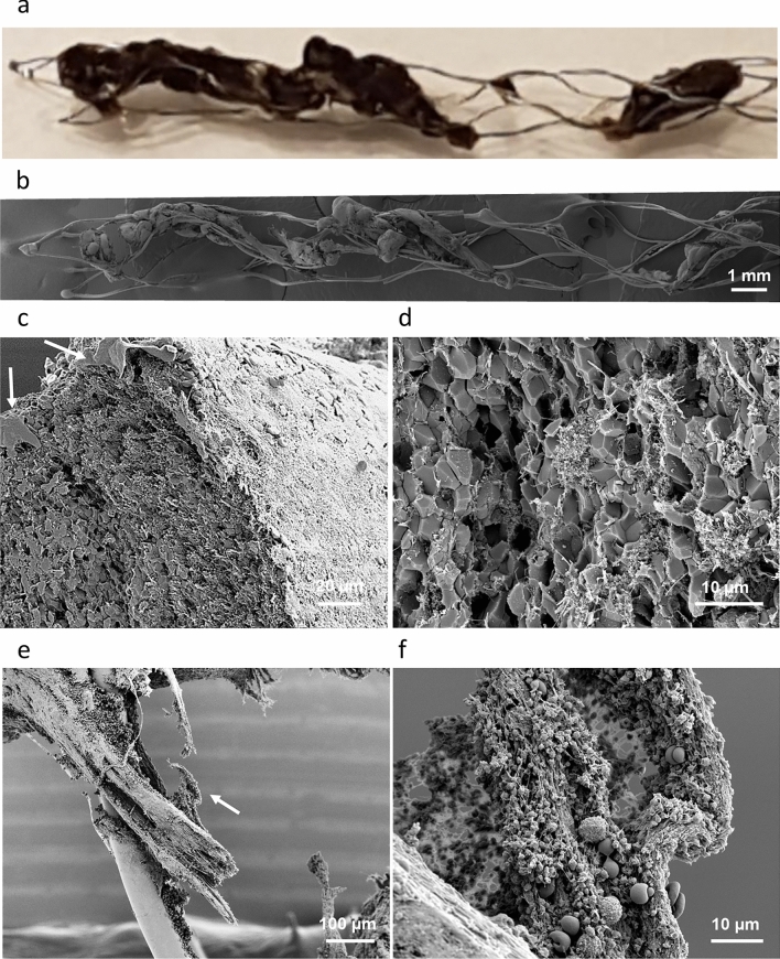
RBCs rich thrombus incorporated onto the stent retriever (Case 2 in Table 1). (a) Optical micrograph. (b) Collage of SEM micrographs. (c) Cross section of a thrombus segment, showing a compact core, porous periphery and fibrin outer layer. Remnants of vascular tissue are visible at the thrombus surface (arrows). (d) High magnification view of the compact core composed of polyhedrocites. (e) Fibrin strings are found in between thrombus segments. (f) White blood cells and platelets are attached to the fibrin strings (higher magnification view of (e), region marked with arrow).

Modalities of RBCs rich thrombus attachment onto the stent. (a) Stent strut protruding through thrombus. (b) Higher magnification view from (a) (dashed rectangle), showing a platelets cap and biconcave RBCs. (c) Thrombus conforming with the stent strut. (d) Higher magnification view from c (region indicated by arrow), illustrating the thrombus contact area with the stent. (e) Bridges of fibrin between the adjacent stent struts. (f) High magnification view from (e) (region indicated by arrow).

Intermediate thrombus incorporated into the stent retriever (Case 4 in Table 1). (a) Optical micrograph. (b, c) SEM view of thrombus on stent. Insert in (c) View of sectioned thrombus and the stent struts at the anchoring site. (d) Fibrin strings wetting the stent surface (indicated by arrows in a, d). (e) Thrombus clutches onto the stent struts. (f) Section through the compact thrombus, showing the contact region with the stent strut (indicated by arrow). (g) Higher magnification view of the thrombus surface at the contact with the stent (indicated by arrow).
Compact structure of an intermediate thrombus. (a) Cross section of thrombus in SEM view. (b) Compact structure of thrombus periphery, with clusters of RBCs encased in a compact matrix of platelets and fibrin. (c) Compact core of thrombus, showing aggregates of polyhedrocites encased in a compact matrix of fibrin and platelets. (d) Higher magnification view from (c), showing the fibrin-platelets matrix.

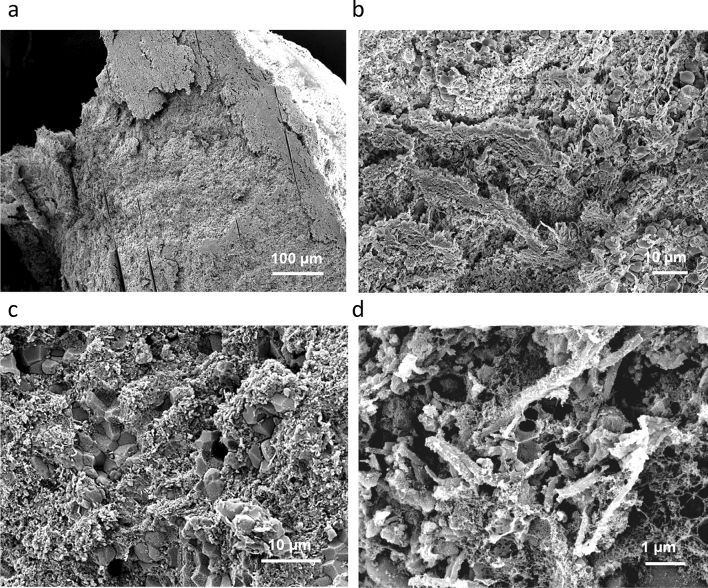
The fibrin rich thrombus integrated in the stent retriever (Case 8 in Table 1). (a) Optical micrograph. (b) Low magnification SEM view. (c) Closer SEM view at the thrombus-stent interface (the stent strut was cut, to allow better viewing). There is no adhesion between the thrombus and the stent strut (arrow).
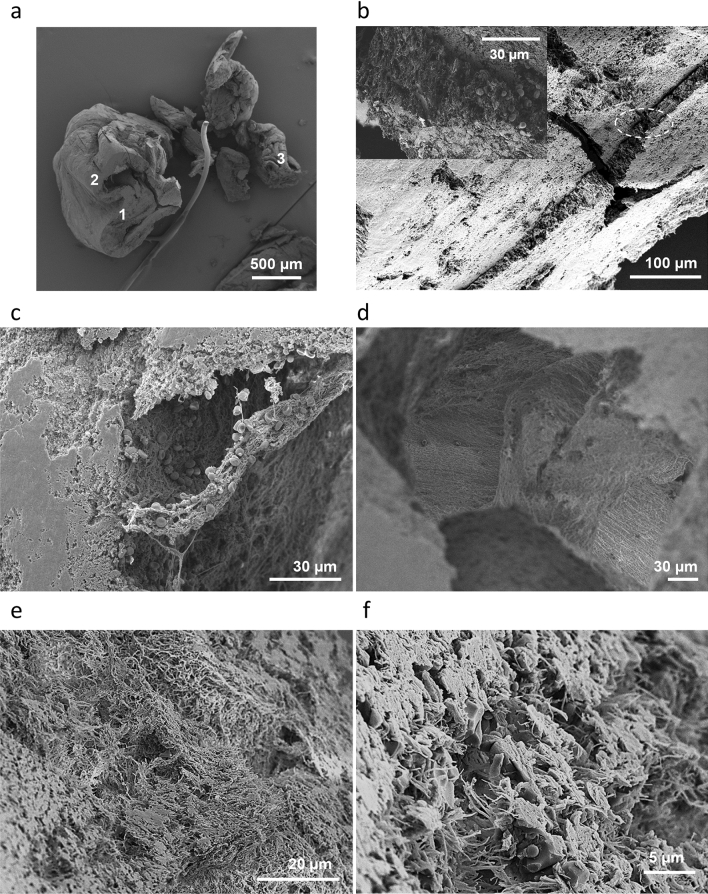
Analysis of fibrin rich thrombus. (a) Cross section of the thrombus. (b) Closer view of the fracture visible on the bigger section of thrombus (marked with “1” in a), and in inset higher magnification of the area marked by the dashed oval. (c), (d) Closer view of the region marked with “2” in (a). (c) Cross section into the wall and scattered red blood cells and white cells inside the cavity. (d) Broad view inside the cavity. (e, f) Cross sections of thrombus segments (marked “3” in a) that were wrapped around the stent strut. (e) Loosely packed fibrin region. (f) Compact region with polyhedral red blood cells, white cells in between the fibrin.
References
Publication types
MeSH terms
Grants and funding
LinkOut - more resources
Full Text Sources
Medical
