Review
. 2022 Nov;38(11):1383-1396.
doi: 10.1007/s12264-022-00860-7. Epub 2022 May 16.

Lighting Up Neural Circuits by Viral Tracing

Affiliations
Review

Lighting Up Neural Circuits by Viral Tracing

Liyao Qiu et al. Neurosci Bull. 2022 Nov.

Abstract

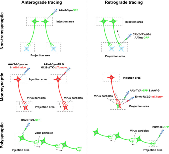
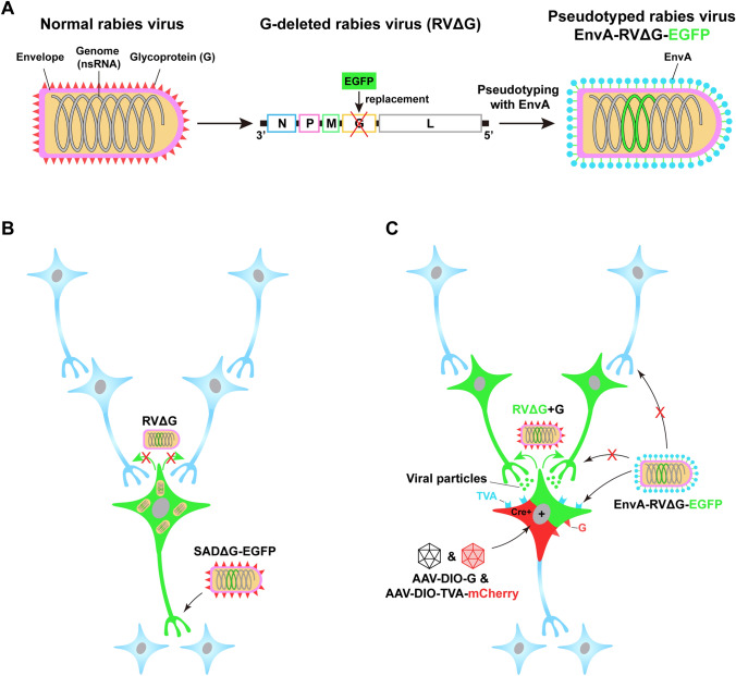
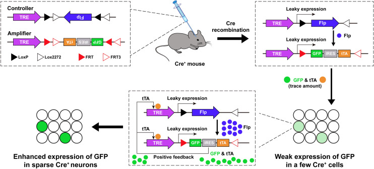
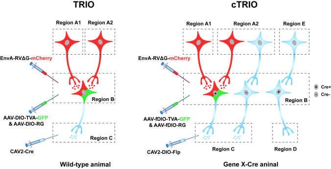
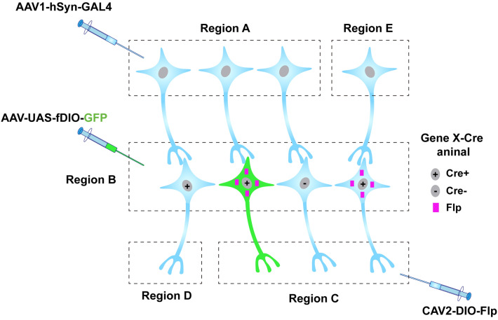
Neurons are highly interwoven to form intricate neural circuits that underlie the diverse functions of the brain. Dissecting the anatomical organization of neural circuits is key to deciphering how the brain processes information, produces thoughts, and instructs behaviors. Over the past decades, recombinant viral vectors have become the most commonly used tracing tools to define circuit architecture. In this review, we introduce the current categories of viral tools and their proper application in circuit tracing. We further discuss some advances in viral tracing strategy and prospective innovations of viral tools for future study.

Keywords: Anterograde; Neural circuit; Retrograde; Transsynaptic; Viral tracing.


Conflict of interest statement

The authors claim that there are no conflict of interest.

Figures

Fig. 1
Fig. 1
Schematic of different viral-tracing strategies. Viral-tracing strategies can be divided into three classes based on their ability to cross synapses: non-transsynaptic (upper), monosynaptic (middle), and polysynaptic (lower). Each class contains both anterograde (left panels) and retrograde (right panels) approaches based on the transport direction of viruses. In anterograde tracing, viruses are often injected into the somal region (injection area), infecting the somas and fully labeling the axonal terminals by expression of fluorescent proteins, thus tracing the terminal regions (projection area). The non-transsynaptic viruses are unable to cross synapses (upper left), whereas the monosynaptic or polysynaptic viruses can transfer to the downstream neurons spanning one (middle left) or multiple synapses (lower left). In non-transsynaptic retrograde tracing, viruses are usually injected into the terminal region, in which they infect the axon terminals and spread retrogradely to the somas (upper right). In monosynaptic or polysynaptic retrograde tracing, viruses are injected into the postsynaptic neuronal areas, are transferred to the presynaptic terminals, and spread retrogradely to the somas (middle right) or to further upstream synaptically-connected neurons (lower right). The neurons that are in both green and red indicate the co-expression of GFP and mCherry, while blue neurons are not infected by viruses (the same convention is used in the following figures). The black arrows indicate the spread direction of viral particles. AAV, adeno-associated virus; AAV1, one subtype of AAV; AAVrg, a retrograde-tracing variant of AAV; CAV, canine adenovirus; hSyn, human Synapsin I; TK, thymidine kinase; HSV, Herpes simplex virus; H129-ΔTK, a TK-deleted anterograde-tracing recombinant of HSV; G, rabies glycoprotein; RVΔG, G-deleted rabies virus; EnvA, avian ASLV type A envelope protein; TVA, avian receptor for EnvA; PRV152, a retrograde-tracing recombinant of the pseudorabies virus.
Fig. 2
Fig. 2
Pseudotyped rabies virus for retrograde tracing. A Engineering rabies virus (RV) by glycoprotein deletion and EnvA pseudotyping. Normal RV (left) contains a negative-strand RNA genome consisting of five genes and an envelope that is coated with the glycoprotein (G), which is coded by one of the five genes. RV can be engineered by replacing the G gene with an enhanced green fluorescent protein (EGFP) and pseudotyping this G-deleted RV with EnvA, the envelope protein of an avian virus (middle and right). B Non-transsynaptic retrograde tracing using SADΔG-EGFP. The recombinant RV SADΔG-EGFP, in which the G-coding gene is deleted, has been coated with G but loses the ability to produce G. This virus can infect axon terminals (shown by the black arrows without red crosses) and retrogradely spread to the somas. It retains the ability to replicate and produce a large amount of virus, thereby enhancing the EGFP fluorescent signal. Unable to synthesize G, however, the newly produced offspring fail to spread to synaptically-connected neurons (shown by the green arrows with red crosses). C Monosynaptic retrograde tracing using EnvA-pseudotyped RV. Due to the absence of endogenous receptors for EnvA, EnvA-RVΔG is unable to infect neurons in the mammalian brain (shown by the black arrows with red crosses). When the EnvA receptor, TVA, is exogenously expressed in the Cre+ neurons via Cre-dependent (DIO) AAV helper vectors, EnvA-RVΔG can selectively infect the TVA-harboring cells (neurons in green plus red), which is shown by the black arrows without red crosses. With the complementation of G in the same cells, the newly generated virus, RVΔG+G regains the ability to transsynaptically spread (shown by the green arrows) to presynaptic neurons (green). Due to the absence of G expression in these presynaptic neurons, however, the virus is unable to further spread out of these cells (blue). The red cross means inability. ns, negative strand; N, nucleoprotein, P, phosphoprotein; M, matrix protein; G, glycoprotein; L, the polymerase of rabies virus; DIO, double-floxed inverse open reading frame; SADΔG, a G-deleted RV strain.
Fig. 3
Fig. 3
A dual-AAV system for sparse labeling of cell-type-specific neurons. Schematic of the design of the dual-AAV system for sparse labeling, comprising a “controller” and an “amplifier” AAV vector (upper left). When these two vectors are mixed and injected into the Cre-expressing mouse, leakage of TRE drives the weak expression of Flp in a few Cre+ neurons. Flp subsequently flips the GFP-IRES-tTA cassettes in the amplifier and drives the low level of GFP and tTA expression in these sparsely-labeled neurons (right panel). The small amount of tTA further binds to the TRE and potentiates the expression of Flp and GFP (lower middle), thereby triggering positive feedback to enhance GFP expression in sparse neurons (lower left). TRE, tetracycline response element; Flp, flippase; IRES, internal ribosome entry site; tTA, tetracycline trans-activator.
Fig. 4
Fig. 4
Viral strategies of TRIO and cTRIO. TRIO combines the application of CAV-2 and RV, thus allowing projection-specific retrograde tracing within three-node neural circuits (from regions A to C via B). cTRIO further combines genetic approaches to allow mapping cell type- and projection-specific neural circuits (from regions A to C via Cre+ cells in B). TRIO, tracing the relationship between input and output; cTRIO, cell-type-specific TRIO; DIO, double-floxed inverse open reading frame; fDIO, Flp-controlled DIO.
Fig. 5
Fig. 5
Intersectional strategies to define and access more specific cell types. With the combination of multiplex recombinase systems, including GAL4-UAS, Cre-LoxP, and Flp-FRT, a more specific subset of neurons can be defined by their input, output, and molecular identity. The schematic shows that neurons expressing Cre and projecting to region C contain Flp. However, only neurons that receive input from region A (shown in green) can express GAL4, which binds to the UAS promoter and initiates the downstream expression of the virus injected into region B. In this case, a specific cell type expressing Cre, which is innervated by certain inputs and projects to putative downstream targets, can be accessed.

Similar articles

Cited by

References

    1. Kristensson K, Olsson Y. Uptake and retrograde axonal transport of peroxidase in hypoglossal neurones. Acta Neuropathol. 1971;19:1–9. - PubMed
    1. Kristensson K, Olsson Y. Retrograde axonal transport of protein. Brain Res. 1971;29:363–365. - PubMed
    1. Gerfen CR, Sawchenko PE. An anterograde neuroanatomical tracing method that shows the detailed morphology of neurons, their axons and terminals: Immunohistochemical localization of an axonally transported plant lectin, Phaseolus vulgaris-leucoagglutinin (PHA-L) Brain Res. 1984;290:219–238. - PMC - PubMed
    1. Glover JC, Petursdottir G, Jansen JK. Fluorescent dextran-amines used as axonal tracers in the nervous system of the chicken embryo. J Neurosci Methods. 1986;18:243–254. - PubMed
    1. Schmued LC, Fallon JH. Fluoro-Gold: a new fluorescent retrograde axonal tracer with numerous unique properties. Brain Res. 1986;377:147–154. - PubMed

LinkOut - more resources