Review
. 2022 Oct;19(6):1691-1704.
doi: 10.1007/s13311-022-01249-0. Epub 2022 May 16.

Major Features of the 2021 WHO Classification of CNS Tumors

Affiliations
Review

Major Features of the 2021 WHO Classification of CNS Tumors

Heather L Smith et al. Neurotherapeutics. 2022 Oct.

Abstract

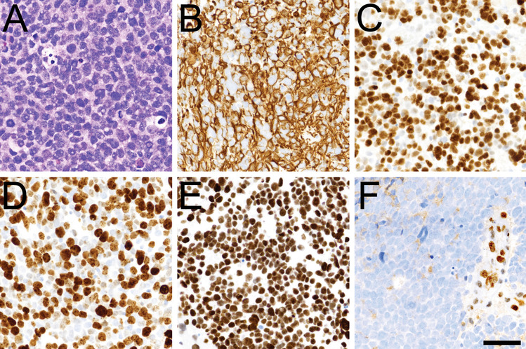
Advances in the understanding of the molecular biology of central nervous system (CNS) tumors prompted a new World Health Organization (WHO) classification scheme in 2021, only 5 years after the prior iteration. The 2016 version was the first to include specific molecular alterations in the diagnoses of a few tumors, but the 2021 system greatly expanded this approach, with over 40 tumor types and subtypes now being defined by their key molecular features. Many tumors have also been reconceptualized into new "supercategories," including adult-type diffuse gliomas, pediatric-type diffuse low- and high-grade gliomas, and circumscribed astrocytic gliomas. Some entirely new tumors are in this scheme, particularly pediatric tumors. Naturally, these changes will impact how CNS tumor patients are diagnosed and treated, including clinical trial enrollment. This review addresses the most clinically relevant changes in the 2021 WHO book, including diffuse and circumscribed gliomas, ependymomas, embryonal tumors, and meningiomas.

Keywords: Astrocytoma; Embryonal; Ependymoma; Glioma; Meningioma; WHO.


Figures

Fig. 1
Fig. 1
Polymorphous low-grade neuroepithelial tumor of the young. In keeping with the “polymorphous” descriptor, polymorphous low-grade neuroepithelial tumor of the young can have a variety of appearances, including that of a diffuse glioma (A), ependymoma (B), and oligodendroglioma (C). These tumors an have abundant mineralization (D) and show abundant CD34 positivity (E) and OLIG2 nuclear staining (F). This particular tumor had a BRAF V600E mutation, and clustered among PLNTYs by DNA methylation profiling
Fig. 2
Fig. 2
Diffuse midline glioma, H3 K27-altered. Diffuse midline gliomas tend to look like most other diffusely infiltrative gliomas (A), including immunopositivity for GFAP (B) and OLIG2 (C). In keeping with their high-grade nature, Ki67 is usually quite high (D). H3 K27M-specific antibody often shows robust nuclear staining (E); those same tumor cells will be weak to negative for H3K27me3, whereas admixed nonneoplastic cells will still be positive (F). Scale bar = 50 microns in all panels
Fig. 3
Fig. 3
Diffuse pediatric-type high-grade glioma, H3-wildtype and IDH-wildtype. This diffuse pediatric-type high-grade glioma, H3 wildtype and IDH-wildtype, looked like a typical diffuse glioma infiltrating into surrounding brain tissue (A); many tumor cells will be Ki67 positive (B). This particular tumor had MYCN amplification and TP53 mutation, mapped to diffuse pediatric-type high-grade glioma, H3 wildtype, and IDH-wildtype by DNA methylation profiling, and had already disseminated throughout the cerebrospinal fluid at the time of clinical presentation. Scale bar = 50 microns in both panels
Fig. 4
Fig. 4
Infant-type hemispheric glioma. Morphologically, infant-type hemispheric gliomas may be indistinguishable from adult-type IDHwt glioblastomas, with palisading necrosis (A), abundant mitoses (B), variable GFAP positivity (C), and elevated Ki67 (D). However, they will not have the same molecular profile as glioblastomas; this tumor had an isolated NTRK fusion, and mapped to infant-type hemispheric glioma by DNA methylation profiling. Scale bar = 250 microns in A, 50 microns in BD
Fig. 5
Fig. 5
Astroblastoma. Astroblastomas can show a variety of morphologies, including heavy sclerosis and mineralization (A). Tumor cells show variable GFAP immunopositivity (B) but are generally positive for OLIG2 (C), epithelial membrane antigen (D), podoplanin (E), and SATB2 (F). Scale bar in F = 100 microns in all panels
Fig. 6
Fig. 6
CNS neuroblastoma, FOXR2-activated. CNS neuroblastoma with FOXR2-activation features sheets of tumor cells with small round nuclei and a high nuclear:cytoplasmic ratio like most other embryonal tumors, although the tumor shown here had more abundant neuropil (A). Like many other new tumor entities, these tumor cells often arrange themselves in a perivascular pattern (B). Most such tumors will also show other high-grade features like necrosis and mitoses, but this one did not. Still, it clearly mapped to CNS neuroblastoma with FOXR2-activation by DNA methylation profiling. Scale bar = 250 microns in A, 100 microns in B
Fig. 7
Fig. 7
CNS tumor with BCOR internal tandem duplication. Many CNS tumors with BCOR internal tandem duplication will look like anaplastic ependymomas, including this one (A, B). But these tumors have a unique molecular profile, and their own DNA methylation pattern. Scale bar = 100 microns

References

    1. Louis DN, Perry A, Reifenberger G, Von Deimling A, Figarella-Branger D, Cavenee WK, et al. The 2016 World Health Organization classification of tumors of the central nervous system: a summary. Acta Neuropathol. 2016;131(6):803–820. doi: 10.1007/s00401-016-1545-1. - DOI - PubMed
    1. Louis DN, Perry A, Wesseling P, Brat DJ, Cree IA, Figarella-Branger D, et al. The 2021 WHO classification of tumors of the central nervous system: a summary. Neuro Oncol. 2021;23(8):1231–1251. doi: 10.1093/neuonc/noab106. - DOI - PMC - PubMed
    1. Horbinski C, Ligon KL, Brastianos P, Huse JT, Venere M, Chang S, et al. The medical necessity of advanced molecular testing in the diagnosis and treatment of brain tumor patients. Neuro Oncol. 2019;21(12):1498–1508. doi: 10.1093/neuonc/noz119. - DOI - PMC - PubMed
    1. Louis DN, Aldape K, Brat DJ, Capper D, Ellison DW, Hawkins C, et al. Announcing cIMPACT-NOW: the Consortium to inform molecular and practical approaches to CNS tumor taxonomy. Acta Neuropathol. 2017;133(1):1–3. doi: 10.1007/s00401-016-1646-x. - DOI - PubMed
    1. Louis DN, Wesseling P, Paulus W, Giannini C, Batchelor TT, Cairncross JG, et al. cIMPACT-NOW update 1: not otherwise specified (NOS) and not elsewhere classified (NEC) Acta Neuropathol. 2018;135(3):481–484. doi: 10.1007/s00401-018-1808-0. - DOI - PubMed

Publication types