. 2022 May 16;13(1):208.
doi: 10.1186/s13287-022-02881-5.

Large-scale engineering of hiPSC-derived nephron sheets and cryopreservation of their progenitors

Affiliations

Large-scale engineering of hiPSC-derived nephron sheets and cryopreservation of their progenitors

Loes E Wiersma et al. Stem Cell Res Ther. .

Abstract

Background: The generation of human induced pluripotent stem cells (hiPSCs) has opened a world of opportunities for stem cell-based therapies in regenerative medicine. Currently, several human kidney organoid protocols are available that generate organoids containing kidney structures. However, these kidney organoids are relatively small ranging up to 0.13 cm2 and therefore contain a small number of nephrons compared to an adult kidney, thus defying the exploration of future use for therapy.

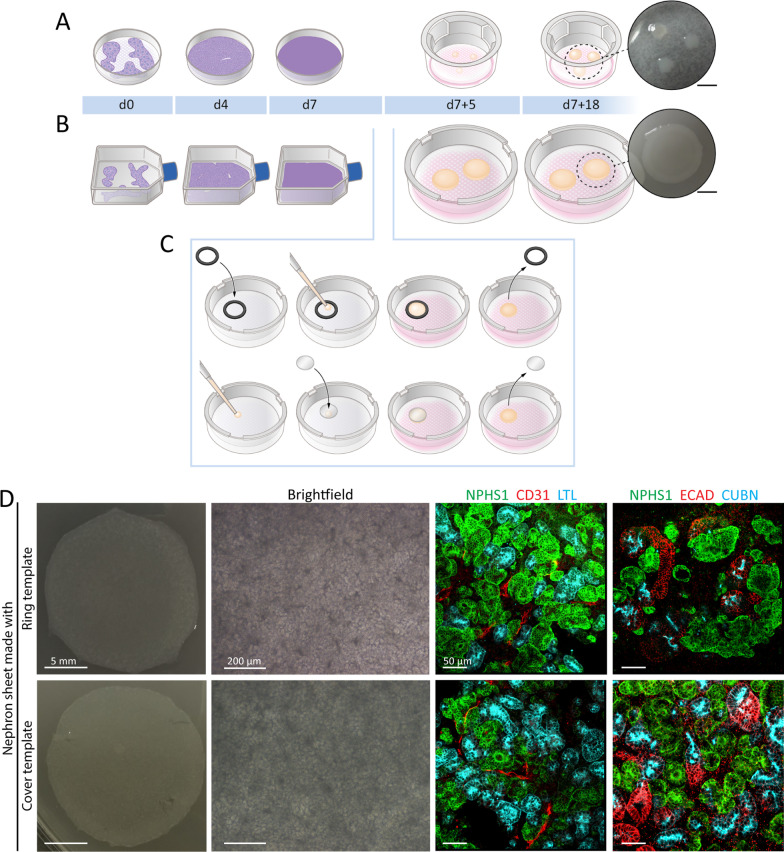
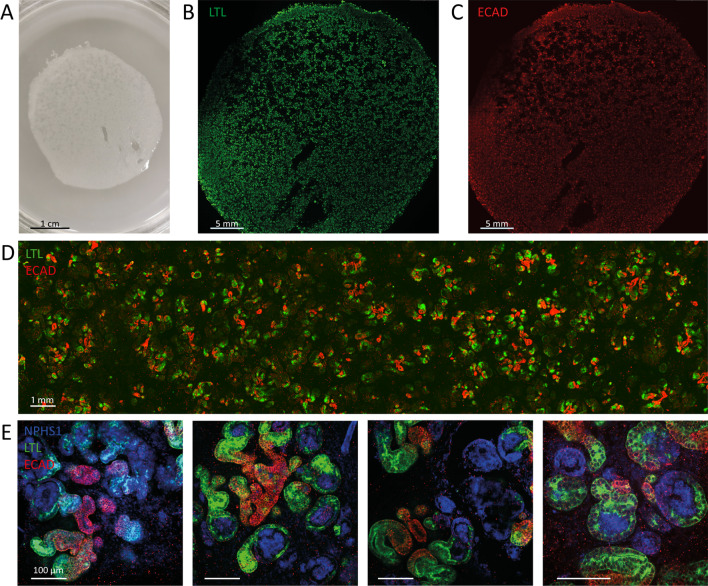
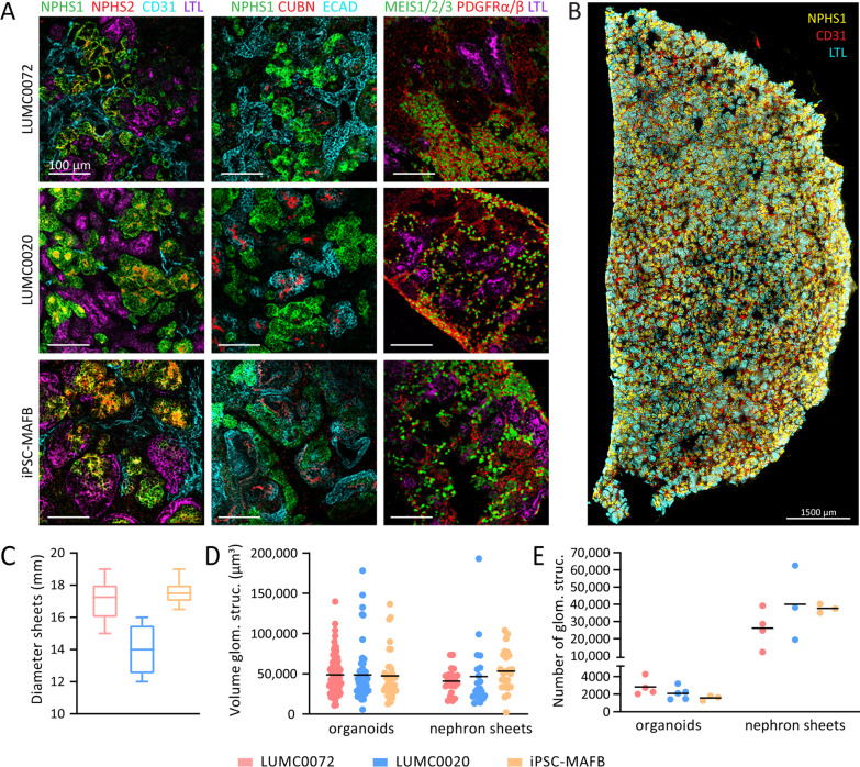
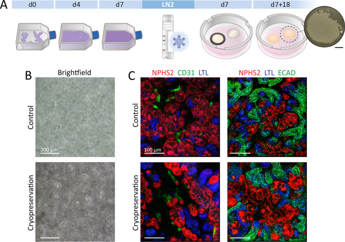
Method: We have developed a scalable, easily accessible, and reproducible protocol to increase the size of the organoid up to a nephron sheet of 2.5 cm2 up to a maximum of 12.6 cm2 containing a magnitude of nephrons.

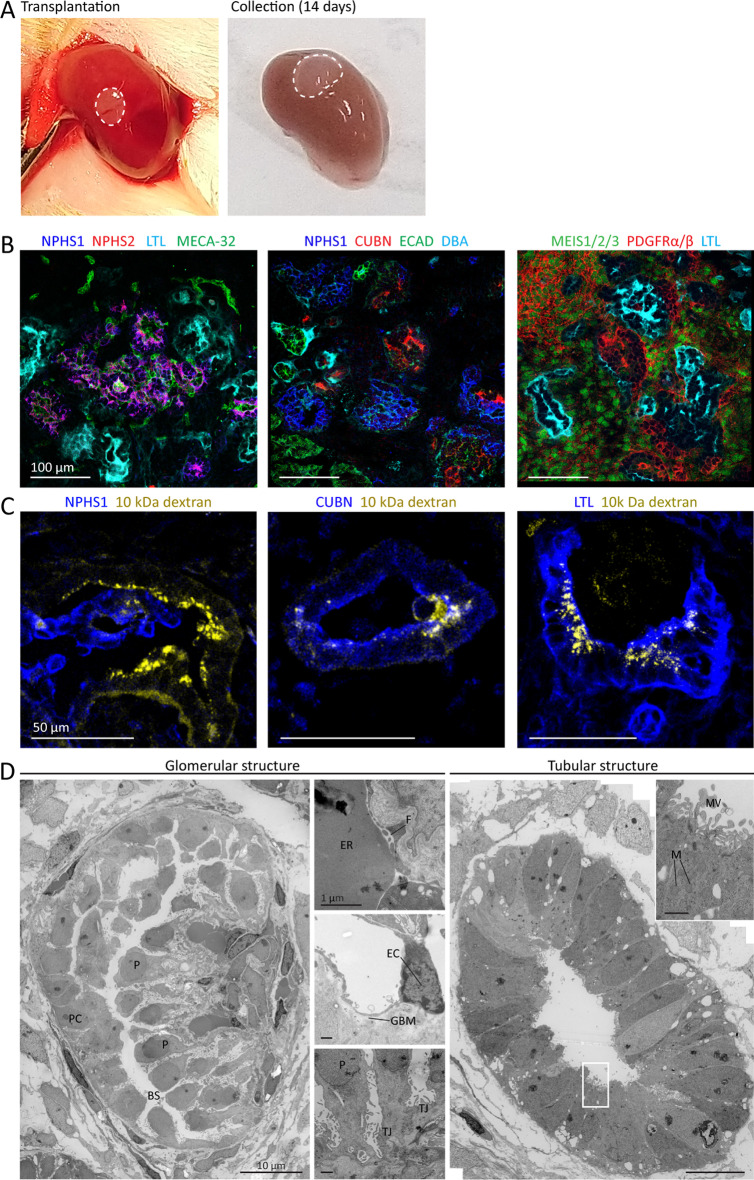
Results: Confocal microscopy showed that the subunits of the nephrons remain evenly distributed throughout the entire sheet and that these tissue sheets can attain ~ 30,000-40,000 glomerular structures. Upon transplantation in immunodeficient mice, such nephron sheets became vascularized and matured. They also show reuptake of injected low-molecular mass dextran molecules in the tubular structures, indicative of glomerular filtration. Furthermore, we developed a protocol for the cryopreservation of intermediate mesoderm cells during the differentiation and demonstrate that these cells can be successfully thawed and recovered to create such tissue sheets.

Conclusion: The scalability of the procedures, and the ability to cryopreserve the cells during differentiation are important steps forward in the translation of these differentiation protocols to future clinical applications such as transplantable auxiliary kidney tissue.

Keywords: Cryopreservation; Engineering; Induced pluripotent stem cells; Kidney organoids; Kidney transplantation; Regenerative medicine; Scale-up.


Conflict of interest statement

The authors declare that they have no competing interests.

Figures

Fig. 1
Fig. 1
Generating hiPSC-derived nephron sheets using a template. A Schematic of the protocol for generating hiPSC-derived kidney organoids. hiPSCs are cultured on multiwell plates as a monolayer until day 7. Cells are dissociated and pipetted as clumps on transwell membranes and cultured until day 7 + 18 (4 mm diameter and 0.13 cm2 surface area). Scalebar inset: 5 mm. B, C Schematic of the protocol for generating hiPSC-derived nephron sheets. hiPSCs are differentiated in T75-culture flasks as a monolayer, followed by dissociation to single cells on day 7. Differentiated cells are either pipetted inside a ring (C, top) or pipetted cells are overlaid with a cover (C, bottom). Nephron sheets are cultured until day 7 + 18 and reach 18 mm diameter with 2.5 cm2 surface area. Scalebar inset: 5 mm. D Images of whole tissue sheet (left) and brightfield (middle), and immunofluorescence (right) of nephron sheets generated with a ring (top) or cover (bottom)
Fig. 2
Fig. 2
Upscaling of nephron sheets to a diameter of at least 4 cm. A Image of large-scale nephron sheet of 12.6 cm2 made with a 4 cm diameter cover template (iPSC-MAFB). B, C Immunofluorescence analysis of the entire nephron sheet shows presence and equal distribution of kidney tubular structures stained for proximal (LTL, B) and distal (ECAD, C) markers. D Section of the large nephron sheet highlights the distribution of tubular structures stained for LTL and ECAD combined. E Detection of nephron structures after immunofluorescence staining for glomerular structures (NPHS1), proximal tubules (LTL), and distal tubules (ECAD)
Fig. 3
Fig. 3
hiPSC-derived nephron sheets reproducibly contain kidney structures and show high number of glomerular structures. A Immunofluorescence analysis for glomerular structures (NPHS1, NPHS2), endothelium (CD31), proximal tubule (LTL, CUBN), distal tubular and collecting duct structures (ECAD) in whole mount nephron sheets, and stromal cells (MEIS1/2/3, PDGFRα/β) in cryosections using LUMC0072, LUMC0020 and iPSC-MAFB (representative images from 3 independent experiments). B Immunofluorescent 3-dimensional overview showing distribution of NPHS1, CD31 and LTL in bisected hiPSC-derived nephron sheet. C Diameter of hiPSC-derived nephron sheets using an 18 mm template in multiple hiPSC lines. (LUMC0072: 32 nephron sheets in 11 independent experiments, LUMC0020: 5 nephron sheets in 3 independent experiments, iPSC-MAFB: 11 nephron sheets in 6 independent experiments). D Volume of individual glomerular structures (µm3) of organoids and nephron sheets. Each dot represents the volume of a single glomerular structure determined in LUMC0072: 7 organoids from 4 independent experiments and 3 nephron sheets from 3 independent experiments; LUMC0020: 6 organoids in 6 independent experiments and 3 nephron sheets in 3 independent experiments; iPSC-MAFB: 4 organoids in 4 independent experiments and 3 nephron sheets in 3 independent experiments. Bar displays average. E Number of glomerular structures in organoids and nephron sheets. Total glomerular number was determined in LUMC0072: 4 organoids from 4 independent experiments and 4 nephron sheets from 4 independent experiments; LUMC0020: 5 organoids in 5 independent experiments and 3 nephron sheets in 3 independent experiments; iPSC-MAFB: 3 organoids in 3 independent experiments and 3 nephron sheets in 3 independent experiments. Bar displays average
Fig. 4
Fig. 4
hiPSC-derived nephron sheet become vascularized and mature upon transplantation. A Macroscopic images of transplanted biopsy of hiPSC-derived nephron sheet (dotted lines, LUMC0072, day 7 + 17) under renal capsule of mice on the day of transplantation (left) and upon collection after 14 days (right). B Immunofluorescence analysis of cryosections demonstrates presence of glomerular structures (NPHS1, NPHS2), mouse endothelial cells (MECA-32), proximal tubules (LTL, CUBN), distal tubules and collecting duct (ECAD, DBA), and stromal cells (MEIS 1/2/3, PDGFRα/β). C Detection of intravenously injected low molecular mass dextran (10 kDa, TRITC labeled) combined with immunofluorescence analysis of glomerular (NPHS1) and proximal tubular (CUBN and LTL) on cryosections demonstrates functional filtration. D Transmission electron micrographs of a glomerular structure and proximal tubule after transplantation. The glomerular structure shows development of a Bowman’s capsule and podocyte orientation towards a capillary (left image), erythrocytes and fenestrae are lining the blood vessel wall (top small image), smoothing of the glomerular basement membrane and endothelial cells are observed (middle small image) and tight junctions connect podocytes (bottom small image). Proximal tubule shows a single layer of epithelial cells, basal orientation of nuclei, mitochondria, and displays brush border with microvilli. P, podocytes; BS, Bowman’s space; PC, parietal cells; ER, erythrocytes; F, fenestrae; TJ, tight junctions; EC, endothelial cell; GBM, glomerular basement membrane; MV, microvilli; N, nuclei; M, mitochondria
Fig. 5
Fig. 5
Cryopreservation of hiPSC-derived nephron sheets. A Schematic of cryopreservation procedure. hiPSCs are differentiated until day 7 of differentiation, dissociated to single cells and cryopreserved. Cells are thawed and differentiation can be continued using both templates for hiPSC-derived nephron sheets. B Brightfield images of nephron sheets without (control) and with cryopreservation (representative image of 4 independent experiments). C Immunofluorescence analysis of glomerular structures (NPHS1, NPHS2), endothelium (CD31), proximal tubules (LTL, CUBN), distal tubular structures (ECAD) in whole mount control and cryopreserved nephron sheets (4 independent experiments)

References

    1. Chronic Kidney Disease Collaboration G.B.D. Global, regional, and national burden of chronic kidney disease, 1990–2017: a systematic analysis for the Global Burden of Disease Study 2017. Lancet 2020;395(10225):709–733. 10.1016/S0140-6736(20)30045-3. - PMC - PubMed
    1. Liyanage T, Ninomiya T, Jha V, et al. Worldwide access to treatment for end-stage kidney disease: a systematic review. Lancet. 2015;385(9981):1975–1982. doi: 10.1016/S0140-6736(14)61601-9. - DOI - PubMed
    1. Thurlow JS, Joshi M, Yan G, et al. Global epidemiology of end-stage kidney disease and disparities in kidney replacement therapy. Am J Nephrol. 2021;52(2):98–107. doi: 10.1159/000514550. - DOI - PMC - PubMed
    1. Himmelfarb J, Vanholder R, Mehrotra R, et al. The current and future landscape of dialysis. Nat Rev Nephrol. 2020;16(10):573–585. doi: 10.1038/s41581-020-0315-4. - DOI - PMC - PubMed
    1. Takahashi K, Tanabe K, Ohnuki M, et al. Induction of pluripotent stem cells from adult human fibroblasts by defined factors. Cell. 2007;131(5):861–872. doi: 10.1016/j.cell.2007.11.019. - DOI - PubMed

Publication types

LinkOut - more resources