Case Reports
. 2022 May 16;18(1):182.
doi: 10.1186/s12917-022-03259-8.

Atypical chorioretinal lesions in Siberian Husky dogs with primary angle-closure glaucoma: a case series

Affiliations
Case Reports

Atypical chorioretinal lesions in Siberian Husky dogs with primary angle-closure glaucoma: a case series

Shin Ae Park et al. BMC Vet Res. .

Abstract

Background: A number of etiologies for different canine chorioretinal lesions have been proved or suggested but some fundic lesions remain unclear in terms of an etiologic diagnosis, treatment options and prognosis. The purpose of this case series is to describe atypical chorioretinal lesions observed in dogs with primary angle-closure glaucoma (PACG).

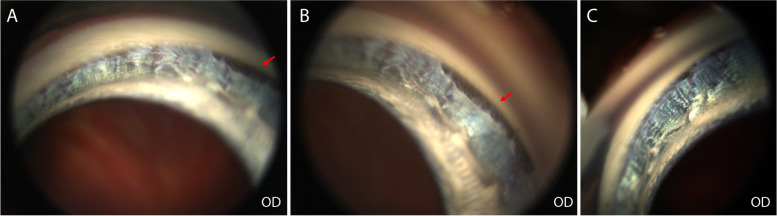
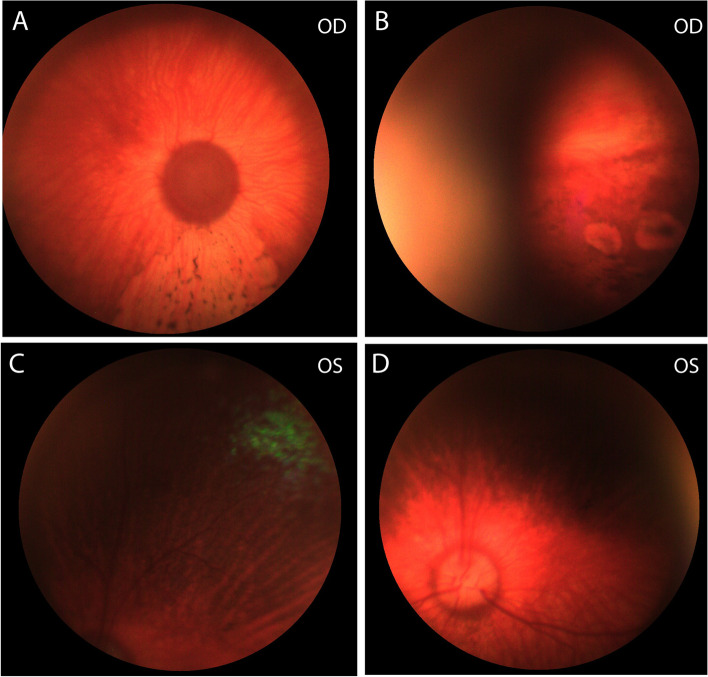
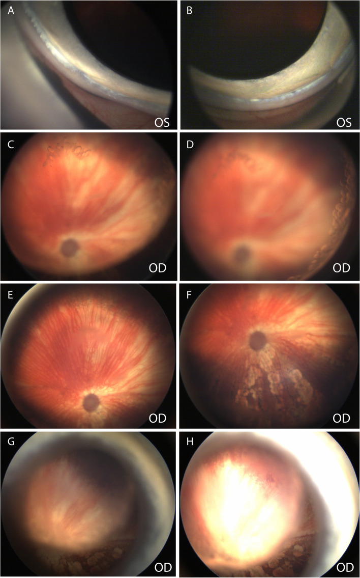
Case presentation: Two spayed-female Siberian Huskies (3- and 4-year-old) and one Siberian Husky/Australian Shepherd mixed breed dog (11-month-old) that had multifocal depigmented retinal lesions and PACG were included.

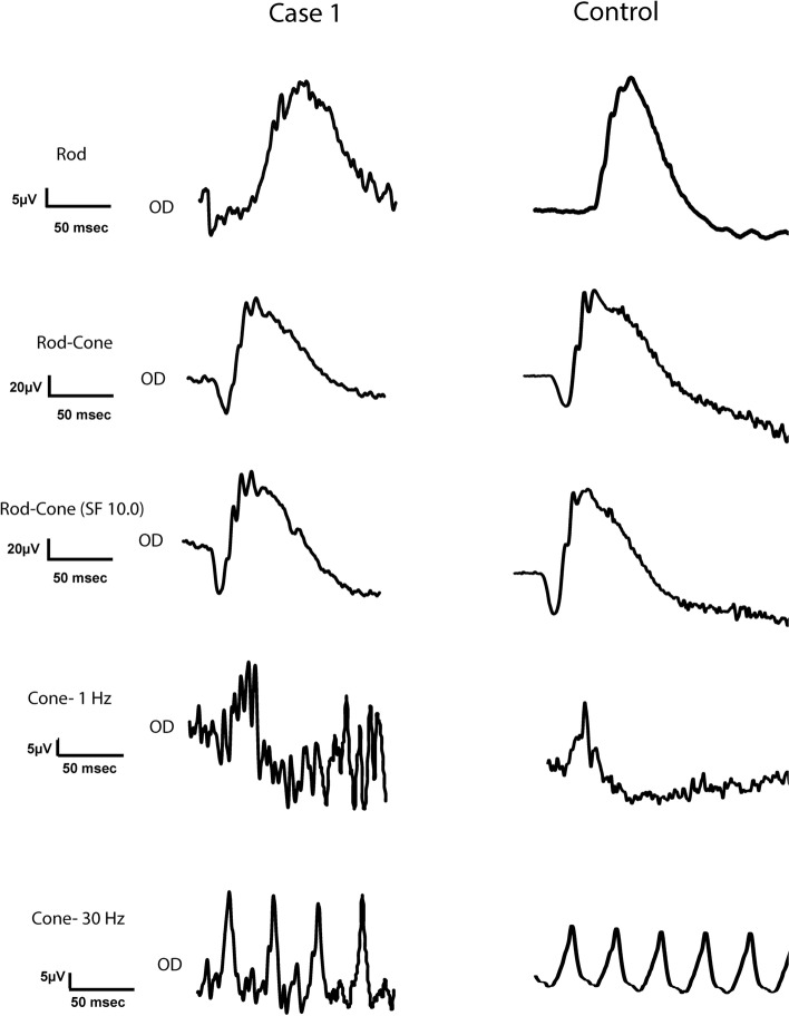
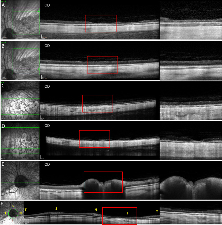
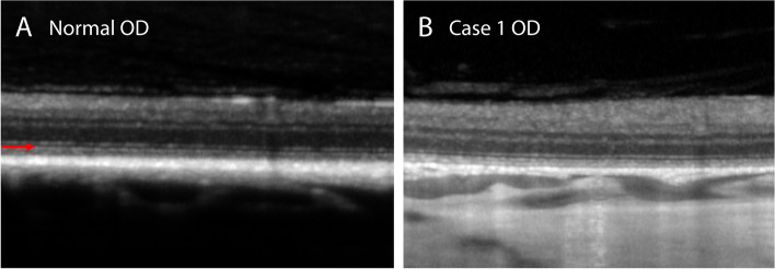
Procedures: Ophthalmic examination, gross, and histopathologic examination findings are described. One of the dogs underwent further clinical diagnostics. Advanced clinical diagnostics on the fellow, presumed to be non-glaucomatous eye of a dog revealed: pectinate ligament dysplasia by gonioscopy, retinal thinning in the depigmented area and wedge shaped retinal thinning with delayed choroidal vascular perfusion by optical coherence tomography, confocal scanning laser ophthalmoscopy, fluorescein and indocyanine green angiography. Quantifiable maze testing for the same eye revealed mild nyctalopia but the full-field electroretinogram showed no generalized decrease of retinal function. Genetic testing for mutations within the retinitis pigmentosa GTPase regulator gene causing X-linked progressive retinal atrophy in Siberian Huskies was negative. Histopathologic evaluations on enucleated eyes in two dogs confirmed goniodysgenesis, PACG with optic nerve head cupping, and diffuse inner retinal atrophy. In addition, segmental profound retinal atrophy, loss of retinal pigment epithelium, and adhesion of the retina to Bruch's membrane was observed and coincided with multifocal depigmented lesions noted on fundic examination.

Conclusions: To our knowledge, this is the first case series with clinical and histopathologic data of chorioretinal lesions, most likely caused by severely impaired choroidal perfusion. Further studies are warranted to elucidate the etiology and pathophysiology, including its possible association with PACG.


Conflict of interest statement

The authors declare that they have no competing interests.

Figures

Fig. 1
Fig. 1
Multifocal irregular shaped depigmented lesions surrounding central pigmented lesions on the inferior fundus in a 4-year-old, spayed female Siberian Husky (case 1, OD). The dog had blue iris and an atapetal fundus OU. Both eyes were symmetrically affected with the multifocal depigmented lesions, but images were obtained from OD only due to corneal edema OS. A and B Images were taken at the first visit. The images are not sharply resolved due to the lack luster corneal surface caused by low tear production. C-E Images were taken at a recheck visit 4 months after the first visit. No grossly notable change in appearance, size and extent of the lesions from the first visit was observed. F The nasal planum had hypopigmentation, which went unchanged throughout the 2-year follow-up period. G, H Close-up images of the fundic lesions with (H) and without (G) a red-free filter show central pigment deposits are located over the choroidal vasculature
Fig. 2
Fig. 2
Narrow iridocorneal angle with moderate pectinate ligament dysplasia was observed in a 4-year-old, spayed female Siberian Husky (case 1, OD). Segmental areas of open angle with normal pectinate ligament (red arrows) were observed in approximately less than 30% of the angle
Fig. 3
Fig. 3
Transit time (A) and number of bumps (B) in a 4-year-old, spayed female Siberian Husky (case1, OD) are higher than normal dogs but lower than the dogs with no rod function with PDE6A mutation in most dim light setting. When the test was repeated with the light setting of 0.02 Lux after a short dark adaptation time (4 mins), the dog in case 1 showed markedly increase in transit time and number of bumps. Time of dark or light adaptation for each light setting is indicated in parentheses in minutes (mins)
Fig. 4
Fig. 4
Normal electroretinography waveforms of a 4-year-old, spayed female, Siberian Husky (case 1, OD) with bilateral multifocal chorioretinal lesions and a 1-year-old normal, intact male, beagle with no ocular disease
Fig. 5
Fig. 5
Optical coherence tomography (OCT) images of A peripheral superonasal, B central superonasal, C inferonasal, D inferior, E horizontal including the optic nerve head, and F peripapillary imaging planes of a 4-year-old, spayed female, Siberian Husky (case 1, OD) showing multifocal partial-to-complete loss of retinal thickness and structure. Note the choroidal thinning and vasculature attenuation (decreased in lumens) in the inferior sections (C and D) compared to the superior sections (A and B). Green horizontal arrows in each infrared confocal scanning laser ophthalmoscopy (cSLO) images indicate the location of the OCT imaging plane. Letters of ‘S’, ‘T’, ‘I’, and ‘N’ in the image indicate superior, temporal, temporal, and nasal quadrant of the fundus. Magnified images of the area within the red rectangle in each image are shown at the right-hand side
Fig. 6
Fig. 6
Optical coherence tomography (OCT) images of A depigmented area, B central pigment deposits, C surrounding area, and D an area of partial loss of retinal layers of a 4-year-old, spayed female, Siberian Husky (case 1, OD). Note the choroidal thinning and vasculature attenuation (decreased in lumen sizes) in the inferior sections (A-C). Green horizontal arrows in each infrared confocal scanning laser ophthalmoscopy (cSLO) image indicate the location of the OCT imaging plane. The red arrow in each cSLO image indicates the location of the vertical green line in the corresponding OCT image. An area of partial loss of retinal layers (D) is observed in the OCT image of the temporal fundus that appears bright in the cSLO image. Magnified images of the area within the red rectangle in each image are shown at the right-hand side
Fig. 7
Fig. 7
Optical coherence tomography (OCT) images show relatively intact ellipsoid zone (red arrow) in a 4-year-old, spayed female, Siberian Husky (case 1, OD) with multifocal depigmented chorioretinal lesions. A For comparison, an OCT image of an intact ellipsoid zone in the superotemporal fundus in a 5-year-old, intact female, mixed-breed dog with no ocular disease (normal OD). B The image was obtained from the superotemporal fundus close to the optic nerve where the retina was relatively well-preserved without depigmented chorioretinal lesions (case 1 OD)
Fig. 8
Fig. 8
A An Infrared (IR) image of the superior fundus; B an IR image of the inferior fundus; C fluorescein angiography (FA, left) and indocyanine green angiography (ICGA, right) images with arterial phase with choroidal filling; D FA (left) and ICGA (right) images with arteriovenous phase; E and F FA (left) image with late venous phase for ICGA and staining phase for FA of a 4-year-old, spayed female, Siberian Husky (case 1, OD). Wedge-shaped lesions were observed in the superonasal fundus with infrared (IR) imaging (A) and inferonasal fundus with fluorescein (left in C and D) and indocyanine green angiography (ICGA, right in C and D). Multifocal hypopigmented areas in the inferior fundus exhibited hyperfluorescence in fluorescein angiography (FA, left in F) but hypofluorescence in ICGA in the late phase (right in F)
Fig. 9
Fig. 9
A Gross image of the enucleated globe showing multifocal reticular shaped depigmented lesions in a 4-year-old, spayed female, Siberian Husky (case 1, OS), the centers of which showed pigment deposits involving the inferior fundus. B A broad, non-perforate, sheet-like band of uveal stroma (asterisk) bridges from the base of the iris to the terminal arborization of Descemet's membrane and extends over the collapsed ciliary cleft. C Histopathologic examination of biopsied nose pad showed variable melanin pigment presence within the basal keratinocyte layers of the epidermis (melanin indicated by arrow). D Histopathologic evaluation OS revealed severe diffuse inner to segmental profound (arrows) retinal atrophy which was consistent with the OCT findings in the fellow eye (OD), which never had a documented IOP spike. E There is multifocal loss of the retinal pigment epithelium and adherence of the neural retina to Bruch's membrane (arrow). The choroid is thin and atrophic. Hematoxylin and eosin (H&E) stain. Scale bars for B, D, and E = 50 μm and for C = 20 μm
Fig. 10
Fig. 10
A Wedge-shaped area without retinal vessels and decreased central RPE pigmentation with focal areas of increased pigmentation surrounding the periphery of noted lesion within the superotemporal fundus of a 3-year old, spayed female, Siberian Husky with glaucoma (case 2, OD). Optic disc cupping and demyelination can be observed. B Two small, circular lesions with lack of pigmentation in the inferotemporal fundus OD but underlying choroidal vessels were observed. C Superotemporal fundus (OS) showing a small area of tapetal fundus. D No significant fundic lesion was observed OS except for slight pallor of the optic nerve head
Fig. 11
Fig. 11
A Gross image of enucleated globe of a 3-year old, spayed female, Siberian Husky (case 2, OD) shows multifocal reticular shaped depigmented lesions, the centers of which showed pigment deposits on the peripheral superior and most of inferior fundus. B A broad, non-perforate, sheet-like band of uveal stroma (asterisk) bridges from the base of the iris to the terminal arborization of Descemet's membrane and extends over the collapsed ciliary cleft. C and D Histopathologic evaluation OD revealed severe diffuse inner to segmental profound retinal atrophy. There is multifocal loss of the retinal pigment epithelium and adherence of the neural retina to Bruch's membrane. The choroid is thin and atrophic. Hematoxylin and eosin (H&E) stain. Scale bars for B and C = 200 μm and for D =100 μm
Fig. 12
Fig. 12
A and B Narrow iridocorneal angle with moderate pectinate ligament dysplasia was observed in an 11-month-old, spayed female Siberian Husky/Australian Shepherd mix (case 3, OS). C Posterior cortical and D equatorial cataracts with vacuoles are observed (OD). E Wedge shaped areas with choroidal vessel attenuation in the superior fundus and F with multifocal irregular shaped depigmented lesions surrounding central pigmented lesions on the inferior fundus OD. A-F Images were obtained at the initial visit. G and H Funduscopic images OD taken 11 months after the initial visit revealed increased area of depigmented lesions. The image is not sharply resolved due to the progressed cataract

References

    1. Narfstrom K, Petersen-Jones SM, Gelatt KN, Gilger BC, Kern TJ, Ames IA. Veterinary ophthalmology. 5. USA: Wiley; 2013. Diseases of the canine ocular fundus; pp. 1303–1392.
    1. Petersen-Jones SM, Komaromy AM. Dog models for blinding inherited retinal dystrophies. Human Gene Ther Clin Dev. 2015;26(1):15–26. doi: 10.1089/humc.2014.155. - DOI - PMC - PubMed
    1. Petersen-Jones SM, Clements PJ, Barnett KC, Sargan DR. Incidence of the gene mutation causal for rod-cone dysplasia type 1 in Irish setters in the UK. J Small Anim Pract. 1995;36(7):310–314. doi: 10.1111/j.1748-5827.1995.tb02934.x. - DOI - PubMed
    1. Winkler PA, Occelli LM, Petersen-Jones SM. Large animal models of inherited retinal degenerations: a review. Cells. 2020;9(4):882. - PMC - PubMed
    1. Genetics Committee of the American College of Veterinary Ophthalmologists . Ocular disorders presumed to be inherited in purebred dogs. 11. American College of Veterinary Ophthalmologists; 2018.

Publication types

LinkOut - more resources