

Emphysema Progression at CT by Deep Learning Predicts Functional Impairment and Mortality: Results from the COPDGene Study
- PMID: 35579519
- PMCID: PMC9434819
- DOI: 10.1148/radiol.213054
Emphysema Progression at CT by Deep Learning Predicts Functional Impairment and Mortality: Results from the COPDGene Study
Abstract
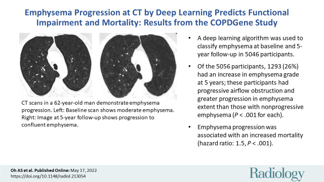
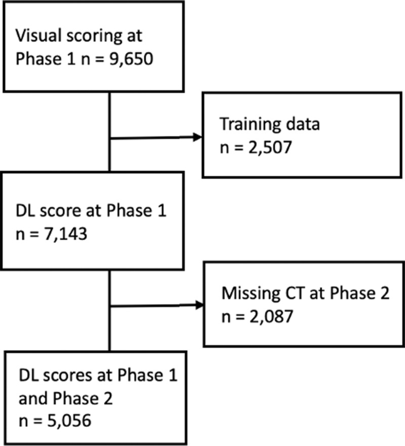
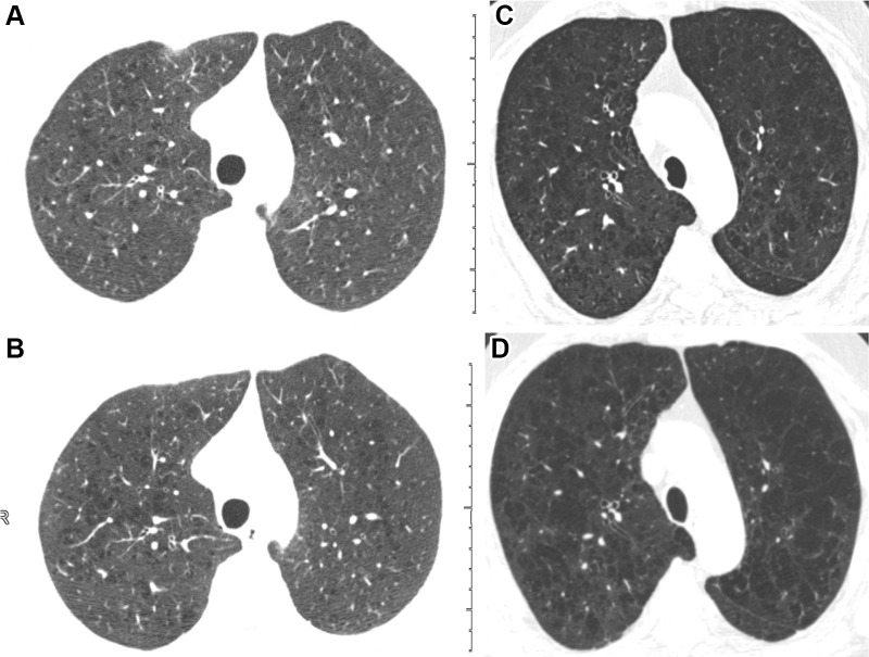
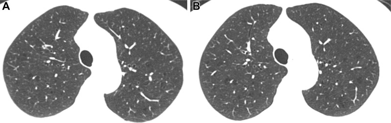
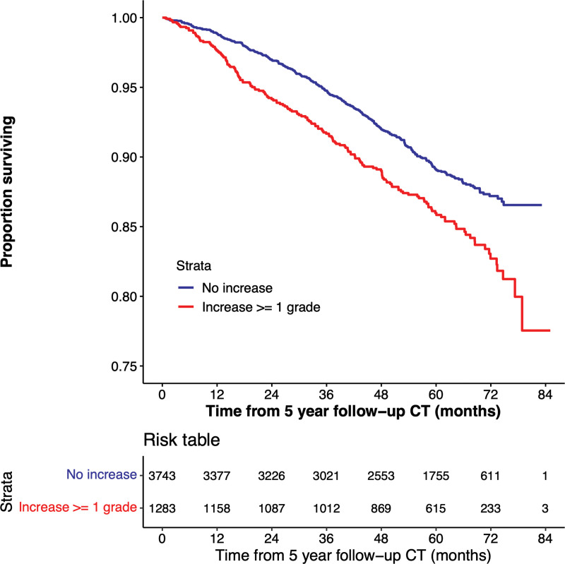
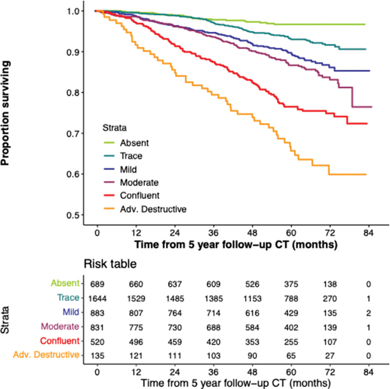
Background Visual assessment remains the standard for evaluating emphysema at CT; however, it is time consuming, is subjective, requires training, and is affected by variability that may limit sensitivity to longitudinal change. Purpose To evaluate the clinical and imaging significance of increasing emphysema severity as graded by a deep learning algorithm on sequential CT scans in cigarette smokers. Materials and Methods A secondary analysis of the prospective Genetic Epidemiology of Chronic Obstructive Pulmonary Disease (COPDGene) study participants was performed and included baseline and 5-year follow-up CT scans from 2007 to 2017. Emphysema was classified automatically according to the Fleischner emphysema grading system at baseline and 5-year follow-up using a deep learning model. Baseline and change in clinical and imaging parameters at 5-year follow-up were compared in participants whose emphysema progressed versus those who did not. Kaplan-Meier analysis and multivariable Cox regression were used to assess the relationship between emphysema score progression and mortality. Results A total of 5056 participants (mean age, 60 years ± 9 [SD]; 2566 men) were evaluated. At 5-year follow-up, 1293 of the 5056 participants (26%) had emphysema progression according to the Fleischner grading system. This group demonstrated progressive airflow obstruction (forced expiratory volume in 1 second [percent predicted]: -3.4 vs -1.8), a greater decline in 6-minute walk distance (-177 m vs -124 m), and greater progression in quantitative emphysema extent (adjusted lung density: -1.4 g/L vs 0.5 g/L; percentage of lung voxels with CT attenuation less than -950 HU: 0.6 vs 0.2) than those with nonprogressive emphysema (P < .001 for each). Multivariable Cox regression analysis showed a higher mortality rate in the group with emphysema progression, with an estimated hazard ratio of 1.5 (95% CI: 1.2, 1.8; P < .001). Conclusion An increase in Fleischner emphysema grade on sequential CT scans using an automated deep learning algorithm was associated with increased functional impairment and increased risk of mortality. ClinicalTrials.gov registration no. NCT00608764 © RSNA, 2022 Online supplemental material is available for this article. See also the editorial by Grenier in this issue.
Conflict of interest statement
Figures
Comment in
-
Deep Learning Assessment of Emphysema Progression at CT Predicts Outcomes.Radiology. 2022 Sep;304(3):680-682. doi: 10.1148/radiol.220627. Epub 2022 May 17. Radiology. 2022. PMID: 35579529 No abstract available.
References
-
- Qaseem A , Wilt TJ , Weinberger SE , et al. . Diagnosis and management of stable chronic obstructive pulmonary disease: a clinical practice guideline update from the American College of Physicians, American College of Chest Physicians, American Thoracic Society, and European Respiratory Society . Ann Intern Med 2011. ; 155 ( 3):179 – 191. - PubMed
-
- Müller NL , Staples CA , Miller RR , Abboud RT . “Density mask”. An objective method to quantitate emphysema using computed tomography . Chest 1988. ; 94 ( 4):782 – 787. - PubMed
-
- Madani A , Zanen J , de Maertelaer V , Gevenois PA . Pulmonary emphysema: objective quantification at multi-detector row CT--comparison with macroscopic and microscopic morphometry . Radiology 2006. ; 238 ( 3):1036 – 1043. - PubMed
-
- Hruban RH , Meziane MA , Zerhouni EA , et al. . High resolution computed tomography of inflation-fixed lungs. Pathologic-radiologic correlation of centrilobular emphysema . Am Rev Respir Dis 1987. ; 136 ( 4):935 – 940. - PubMed
Publication types
MeSH terms
Associated data
Grants and funding
LinkOut - more resources
Full Text Sources
Medical
