. 2022 Sep 7;14(661):eabm7621.
doi: 10.1126/scitranslmed.abm7621. Epub 2022 Sep 7.

The adenosine analog prodrug ATV006 is orally bioavailable and has preclinical efficacy against parental SARS-CoV-2 and variants

Affiliations

The adenosine analog prodrug ATV006 is orally bioavailable and has preclinical efficacy against parental SARS-CoV-2 and variants

Liu Cao et al. Sci Transl Med. .

Abstract

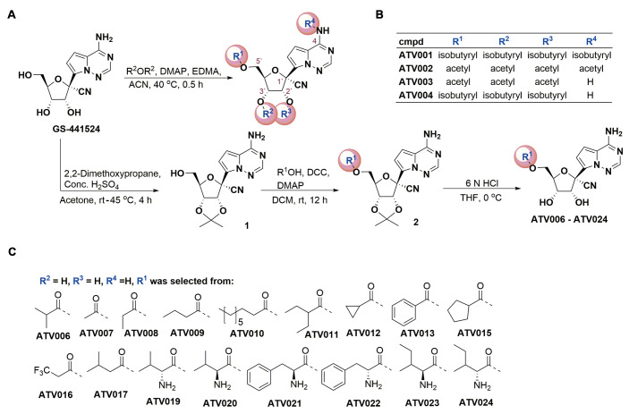
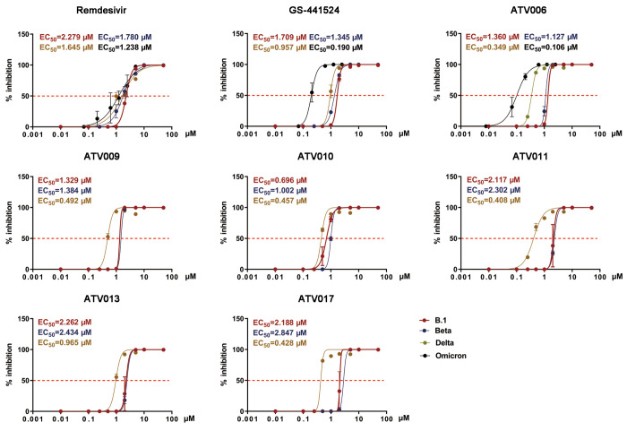
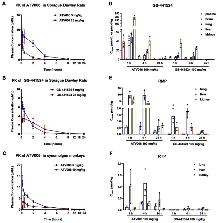
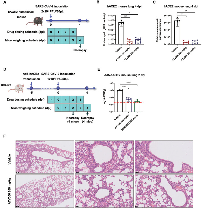
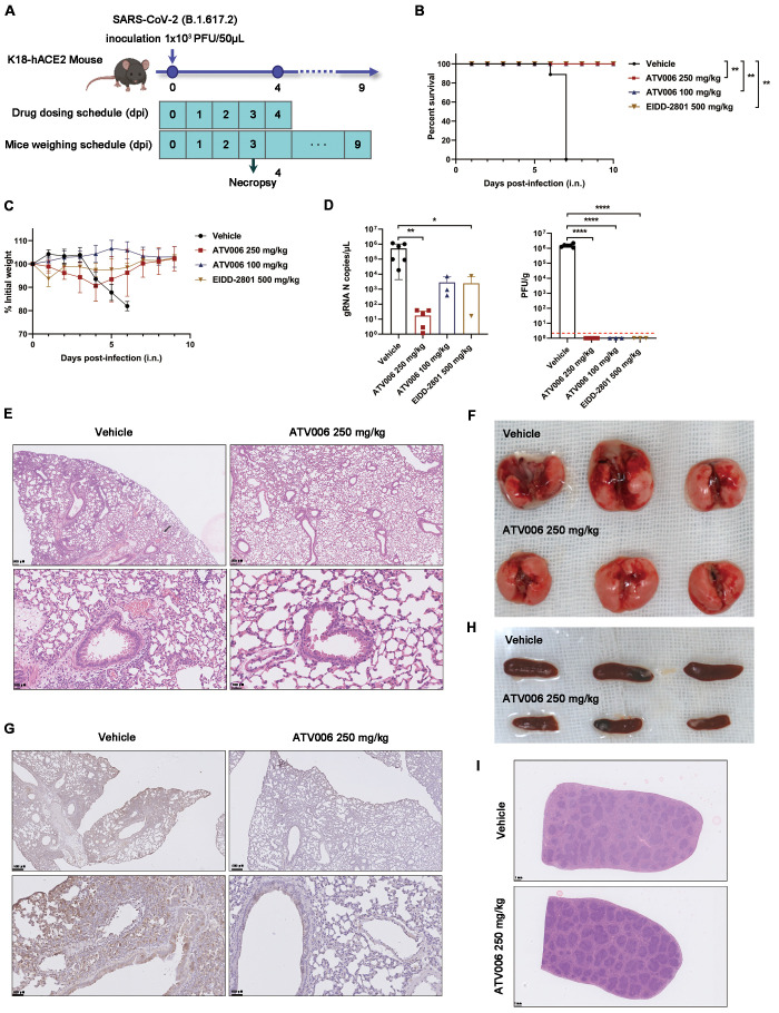
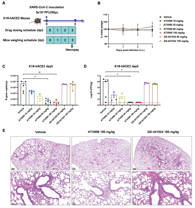
Severe acute respiratory syndrome coronavirus 2 (SARS-CoV-2), the virus driving the ongoing coronavirus disease 2019 (COVID-19) pandemic, continues to rapidly evolve. Because of the limited efficacy of vaccination in prevention of SARS-CoV-2 transmission and continuous emergence of variants of concern (VOCs), orally bioavailable and broadly efficacious antiviral drugs are urgently needed. Previously, we showed that the parent nucleoside of remdesivir, GS-441524, has potent anti-SARS-CoV-2 activity. Here, we report that esterification of the 5'-hydroxyl moieties of GS-441524 markedly improved antiviral potency. This 5'-hydroxyl-isobutyryl prodrug, ATV006, demonstrated excellent oral bioavailability in rats and cynomolgus monkeys and exhibited potent antiviral efficacy against different SARS-CoV-2 VOCs in vitro and in three mouse models. Oral administration of ATV006 reduced viral loads and alleviated lung damage when administered prophylactically and therapeutically to K18-hACE2 mice challenged with the Delta variant of SARS-CoV-2. These data indicate that ATV006 represents a promising oral antiviral drug candidate for SARS-CoV-2.


Figures

Fig. 1.
Fig. 1.. The chemical structure and synthesis of GS-441524-derived prodrugs.
(A) The synthetic process of GS-441524 derivatives is shown. R2OR2, Acid anhydrides with carbonyl of R2; rt, room temperature; DCC, Dicyclohexylcarbodiimide; THF, Tetrahydrofuran. (B) Compounds (cmpd) ATV001 to ATV004 were synthesized from GS-441524 by one-step acylation reactions with related acid anhydride in the presence of 4-dimethylaminopyridine (DMAP) and ethylene dimethacrylate (EDMA). (C) For the synthesis of ATV006-024, 2′,3′-hydroxyl moieties of GS-441524 were protected with acetonide by using 2,2-dimethoxypropane in the presence of sulfuric acid (H2SO4). Different aliphatic acids, aromatic acids, or amino acids were reacted with intermediate 1 to product the corresponding esters by DCC/DMAP-mediated condensation, respectively. The subsequent hydrolysis reaction of compound 2 with 6N hydrochloric acid (HCl) yielded desired compounds ATV006 to ATV024.
Fig. 2.
Fig. 2.. GS-441524 derivatives exhibit antiviral activity against SARS-CoV-2 variants in vitro.
Vero E6 cells were infected with different strains of SARS-CoV-2(B.1, Beta, Delta and Omicron) at a multiplicity of infection (MOI) of 0.05 and treated with dilutions of the indicated compounds for 48 hours. Viral yield in the cell supernatant was then quantified by qRT-PCR [% inhibition= (viral RNA copies of treatment group / viral RNA copies of placebo control group) × 100]. The values of the concentration for 50% of maximal effect (EC50) for each compound are shown above each plot and indicated with the red dashed line.
Fig. 3.
Fig. 3.. Pharmacokinetic profile of ATV006 and GS-441524 in Sprague Dawley rats and cynomolgus monkeys.
(A) A time-plasma concentration curve is shown for the nucleoside GS-441524 following a single IV administration of ATV006 (5 mg/kg) or oral administration (25 mg/kg) to Sprague Dawley rats (n = 3, mean ± SD). (B) A time-plasma concentration curve is shown for the nucleoside GS-441524 following a single IV administration of GS-441524 (5 mg/kg) or oral administration (25 mg/kg) to Sprague Dawley rats (n = 3, mean ± SD). (C) A time-plasma concentration curve is shown for the nucleoside GS-441524 following a single IV administration of ATV006 (5 mg/kg) or oral administration (10 mg/kg) to cynomolgus monkeys (n = 3, mean ± SD). (D to F) The tissue distribution of GS-441524 (D), remdesivir nucleotide monophosphate (RMP) (E), and remdesivir nucleotide triphosphate (RTP) (F) is shown for plasma, brain, lung, liver, and kidney after oral administration of 100 mg/kg ATV006 or GS-441524 to Sprague Dawley rats (n = 3, mean ± SD).
Fig. 4.
Fig. 4.
ATV006 treatment reduces viral load and prevents lung pathology in hACE2 knock-in and Ad5-hACE2 mouse models. (A) Experimental timeline for SARS-CoV-2 infection in hACE2 humanized mice. Mice were intranasally inoculated with the B.1 strain of SARS-CoV-2 (2 × 10⁵ PFU per mouse) and were treated with vehicle (control), ATV006 (250 mg/kg, orally, once daily), or ATV006 (500 mg/kg, orally, once daily) starting at the time of infection (n=6 mice per group). (B and C) Viral load was measured in the lungs at 4 dpi by qRT-PCR analysis of nucleocapsid (N) gRNA (B) and sgRNA (C). The limit of detection of qRT-PCR was 0.5 copies/μL. (D) Experimental timeline for SARS-CoV-2 infection in Ad5-hACE2 mice. Ad5-hACE2-transduced mice infected with B.1 strain of SARS-CoV-2 (1 × 10⁵ PFU per mouse) were treated with vehicle (control), ATV006 (250 mg/kg, orally, once daily), or EIDD-2801 (500 mg/kg, orally, once daily) (n=8 mice per group). (E) Viral titers were measured in the lungs of Ad5-hACE2 mice at 2 dpi by focus forming assay (FFA) and reported as focus-forming units (FFU) per gram of lung tissue. The red dashed line in (E) indicates the limit of detection. (F) Histopathology analysis is shown for lungs isolated at 4 dpi from SARS-CoV-2 infected Ad5-hACE2 mice treated with vehicle or ATV006 (250 mg/kg). Data are presented as mean ± SD. Statistical analysis was conducted using one-way ANOVA with Dunnett’s correction for multiple comparisons (lung viral sgRNA and lung virus titer) or Kruskal-Wallis test with Dunn's correction for multiple comparisons (lung viral gRNA). ∗p ≤ 0.05; ∗∗p ≤ 0.005; ∗∗∗p ≤ 0.0005; ∗∗∗∗p ≤ 0.0001.
Fig. 5.
Fig. 5.. Prophylactic ATV006 treatment is efficacious against SARS-CoV-2 in K18-hACE2 mice.
(A) The experimental timeline is shown. K18-hACE2 mice were intranasally inoculated with the SARS-CoV-2 Delta variant (1 × 10³ PFU per mouse) and treated with vehicle (control, n=11), ATV006 (250 mg/kg, orally, once daily, n=11), ATV006 (100 mg/kg, orally, once daily, n=8) or EIDD-2801 (500 mg/kg, orally, once daily, n=8). (B) The survival curve is shown. (C) Change in body weight was measured over time post infection. (D) Viral load from lung tissue at 3 dpi was analyzed by qRT-PCR and plaque assay. The detection limit of qRT-PCR was 0.5 copies/μL. The red dashed line in (D, right panel) indicates the limit of detection for the plaque assay. (E to G) Histopathology (E), gross pathology (F), and immunohistochemistry detection of SARS-CoV-2 S protein (G) are shown for lungs isolated from the indicated treatment groups. S protein is denoted by brown staining in (G). (H and I) Gross pathology (H) and histopathology (I) are shown for spleens isolated from the indicated treatment groups. Data in (C and D) are presented as mean ± SD. Statistical analysis was conducted using a Log-Rank test (survival), a one-way ANOVA with Dunnett’s correction for multiple comparisons (lung titer), or a Kruskal-Wallis test with Dunn's correction for multiple comparisons (lung viral RNA). ∗p ≤ 0.05; ∗∗p ≤ 0.005; ∗∗∗∗p ≤ 0.0001.
Fig. 6.
Fig. 6.. Therapeutic ATV006 treatment is efficacious against SARS-CoV-2 in K18-hACE2 mice.
(A) The experimental timeline is shown. K18-hACE2 mice were intranasally inoculated with the SARS-CoV-2 Delta variant (5 × 10² PFU per mouse) and were treated with vehicle (control, n=5), ATV006 (at 10, 30, 80, or 150 mg/kg orally, BID, n=5), or GS-441524 (at 80 or 150 mg/kg orally, BID, n=5). (B) Change in body weight was measured over time post infection. (C and D) Viral titers from lungs tissue at 3 dpi were analyzed by qRT-PCR (C) and focus-forming assay (FFA) (D). The detection limit of qRT-PCR was 0.5 copies/μL. The red dashed line in (D, right panel) indicates the limit of detection for the FFA. (E) Histopathology is shown for lungs isolated from mice in the indicated treatment groups. Data in (B to D) are presented as mean ± SD. Statistical analysis was conducted using Kruskal-Wallis tests with Dunn's correction for multiple comparisons (lung viral RNA, lung titer). ∗p ≤ 0.05; ∗∗p ≤ 0.005.

References

    1. World Health Organization (WHO), (2022), WHO Coronavirus (COVID-19) Dashboard. Retrieved on May 3,2022 from https://covid19.who.int.
    1. Chen Y., Liu Q., Guo D., Emerging coronaviruses: Genome structure, replication, and pathogenesis. J. Med. Virol. 92, 2249 (2020). 10.1002/jmv.26234 - DOI - PMC - PubMed
    1. Perlman S., Netland J., Coronaviruses post-SARS: Update on replication and pathogenesis. Nat. Rev. Microbiol. 7, 439–450 (2009). 10.1038/nrmicro2147 - DOI - PMC - PubMed
    1. Al-Tawfiq J. A., Memish Z. A., Middle East respiratory syndrome coronavirus: Transmission and phylogenetic evolution. Trends Microbiol. 22, 573–579 (2014). 10.1016/j.tim.2014.08.001 - DOI - PMC - PubMed
    1. Robson F., Khan K. S., Le T. K., Paris C., Demirbag S., Barfuss P., Rocchi P., Ng W. L., Coronavirus RNA Proofreading: Molecular Basis and Therapeutic Targeting. Mol. Cell 79, 710–727 (2020). 10.1016/j.molcel.2020.07.027 - DOI - PMC - PubMed

Publication types

Substances

Supplementary concepts