. 2022 Jul 1;132(13):e159814.
doi: 10.1172/JCI159814.

A translatable RNAi-driven gene therapy silences PMP22/Pmp22 genes and improves neuropathy in CMT1A mice

Affiliations

A translatable RNAi-driven gene therapy silences PMP22/Pmp22 genes and improves neuropathy in CMT1A mice

Marina Stavrou et al. J Clin Invest. .

Abstract

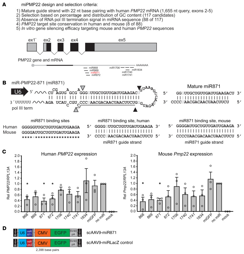
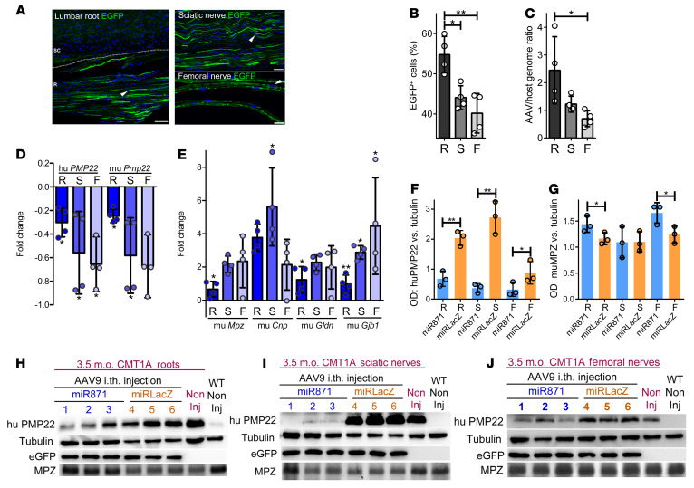
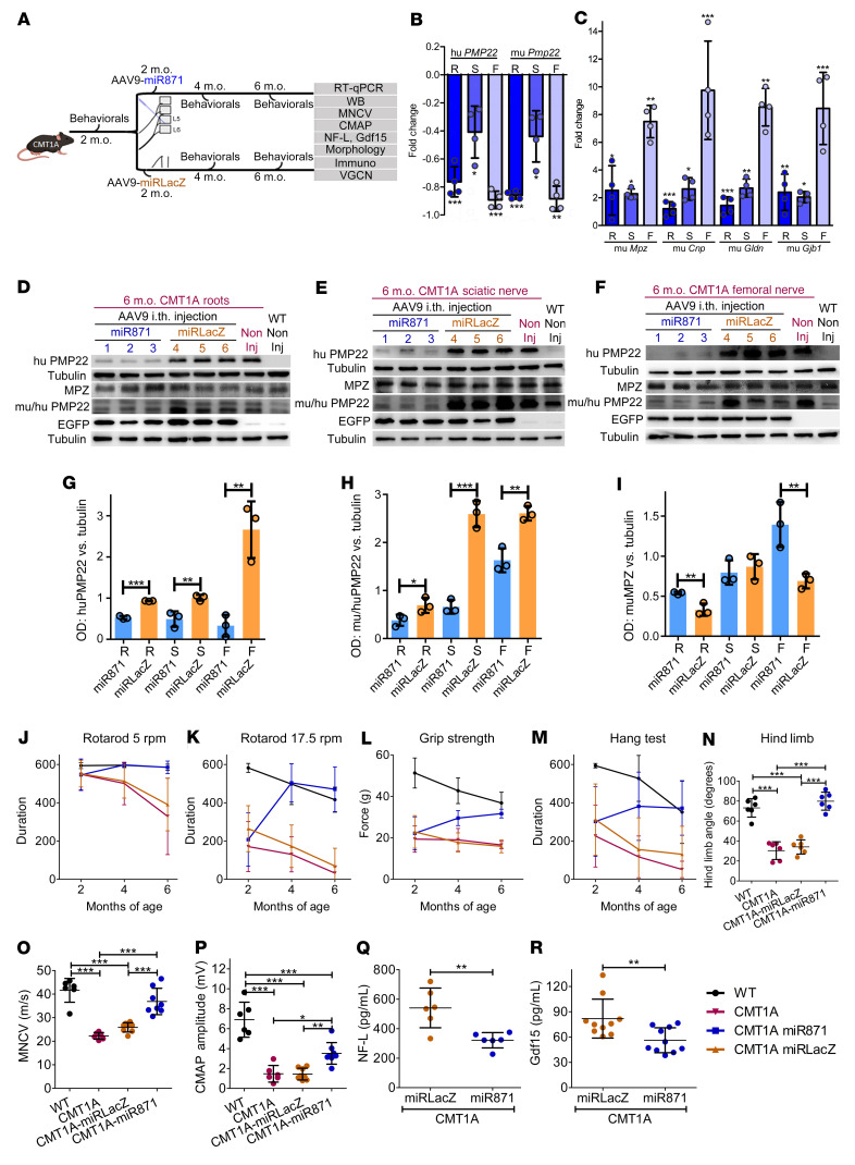
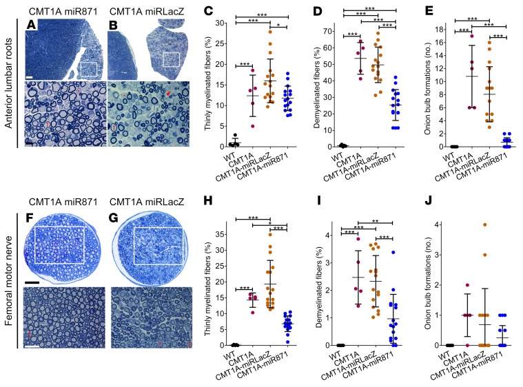
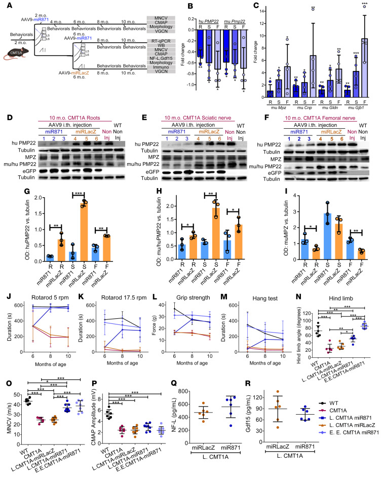
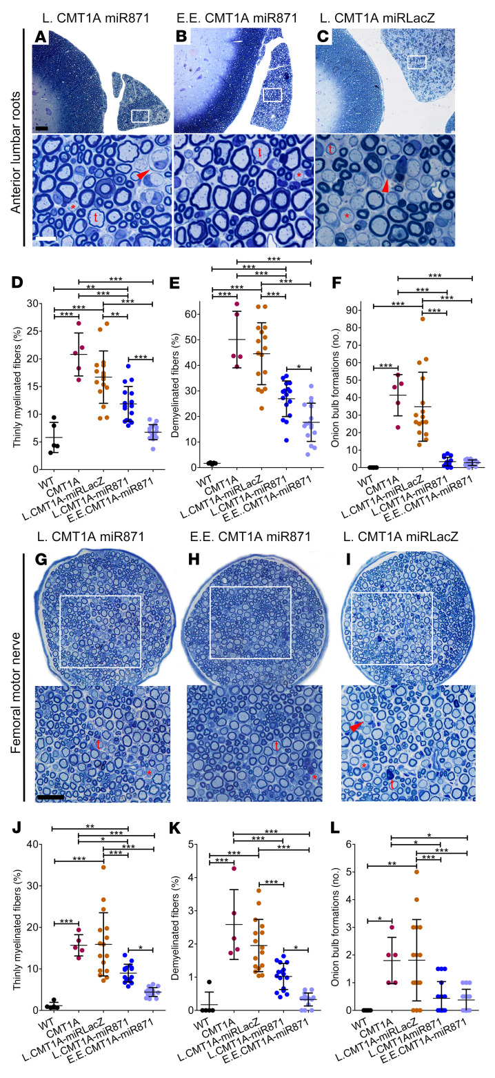
Charcot-Marie-Tooth disease type 1A (CMT1A), the most common inherited demyelinating peripheral neuropathy, is caused by PMP22 gene duplication. Overexpression of WT PMP22 in Schwann cells destabilizes the myelin sheath, leading to demyelination and ultimately to secondary axonal loss and disability. No treatments currently exist that modify the disease course. The most direct route to CMT1A therapy will involve reducing PMP22 to normal levels. To accomplish this, we developed a gene therapy strategy to reduce PMP22 using artificial miRNAs targeting human PMP22 and mouse Pmp22 mRNAs. Our lead therapeutic miRNA, miR871, was packaged into an adeno-associated virus 9 (AAV9) vector and delivered by lumbar intrathecal injection into C61-het mice, a model of CMT1A. AAV9-miR871 efficiently transduced Schwann cells in C61-het peripheral nerves and reduced human and mouse PMP22 mRNA and protein levels. Treatment at early and late stages of the disease significantly improved multiple functional outcome measures and nerve conduction velocities. Furthermore, myelin pathology in lumbar roots and femoral motor nerves was ameliorated. The treated mice also showed reductions in circulating biomarkers of CMT1A. Taken together, our data demonstrate that AAV9-miR871-driven silencing of PMP22 rescues a CMT1A model and provides proof of principle for treating CMT1A using a translatable gene therapy approach.

Keywords: Gene therapy; Mouse models; Neurodegeneration; Neuroscience; Therapeutics.


Conflict of interest statement

Conflict of interest: The sequences and methods described here were included in a provisional patent application filed on December 1, 2020 (PCT/US21/61177). SQH, KAK, and MS are listed as inventors. HZ has served on scientific advisory boards and/or as a consultant for Abbvie, Alector, Annexon, Artery Therapeutics, AZTherapies, Cognition Therapeutics (CogRx), Denali Therapeutics, Eisai Company, NervGen Pharma, Novo Nordisk, Pinteon Therapeutics, Red Abbey Labs, Passage Bio, Roche, Samumed, Siemens Healthineers, Triplet Therapeutics, and Wave; has given lectures at symposia sponsored by Cellectricon, Fujirebio, Alzecure, Biogen, and Roche; and is a co-founder of Brain Biomarker Solutions in Gothenburg AB (BBS), which is a part of the GU Ventures Incubator Program.

Figures

Figure 1
Figure 1. Design and in vitro screen of artificial miRNAs targeting PMP22.
(A) Full-length PMP22 mRNA was transcribed from 5 exons (ex), producing 2 major transcripts with identical ORFs (black shading). We designed 8 candidate miRNAs to equally target both human PMP22 and murine Pmp22 mRNAs. (B) Gray and black arrowheads show miR871 cut sites by Drosha and Dicer, respectively. dsRNAs form G:U wobble base pairs (indicated by gray shading). Underlined sequence represents the mature miR871 antisense guide strand. Bottom panel shows alignment of the miR871 binding site on human PMP22 and murine Pmp22 mRNAs. Gray asterisk indicates a G:A mismatch at the miR871 binding site, but each nucleotide at this location can form 2 hydrogen bonds with the miR871 guide strand as a G:U wobble (human) or A:U wobble (mouse). (C) RT-qPCR was performed to measure in vitro human PMP22 or murine Pmp22 silencing by the indicated miRPMP22s (n = 3/group). Gene expression was normalized to human RPL13A. *P < 0.05, by unpaired, 1-tailed Student’s t test. Values represent the mean ± SEM. Rel, relative. (D) Schematic of scAAV9, which was used to deliver miR871 or miRLacZ expression cassettes in vivo. The U6 promoter drives the transcription of miR871 or miRLacZ, and the CMV promoter drives the EGFP gene with the SV40 polyadenylation sequence.
Figure 2
Figure 2. In vivo assessment of AAV9-miR transduction in PNS tissues and validation of AAV9-miR871 silencing efficiency in a CMT1A mouse model 6 weeks after injection.
(A) Lumbar spinal roots and sciatic nerve sections as well as teased femoral nerve fibers showing EGFP autofluorescence in SCs and axons. Arrowheads indicate examples of EGFP+ nuclei. Scale bars: 60 μm (lumbar root and sciatic nerve) and 20 μm (femoral nerve). (B) Quantification of EGFP-expressing PNS cells (n = 4/group). (C) VGCNs (n = 4/group) confirmed peripheral nerve transduction. RT-qPCR analysis of (D) Human (hu) PMP22 and murine (mu) Pmp22 and of murine (E) Mpz, Cnp, Gldn, and Gjb1 gene expression (n = 3/group). Fold changes in relative mRNA expression of CMT1A-AAV9-miR871 were calculated in comparison with expression levels in CMT1A-AAV9-miRLacZ mice. All samples were normalized to endogenous Gapdh. Quantification of (F) human PMP22 and (G) murine MPZ Western blot protein ODs, normalized to tubulin (Tub), in CMT1A-AAV9-miR871 and CMT1A-AAV9-miRLacZ mice in lumbar roots, sciatic nerves, and femoral nerves. Western blots showing human PMP22, murine tubulin, EGFP, and murine MPZ protein levels in (H) roots, (I) sciatic nerves, and (J) femoral nerves. Values represent the mean ± SD. *P < 0.05 and **P < 0.01, by 1-way ANOVA with Tukey’s multiple-comparison test. R, lumbar roots; S, sciatic nerves; F, femoral nerves; i.th., intrathecal; m.o., months old; Non Inj, noninjected.
Figure 3
Figure 3. Efficient PMP22/Pmp22 silencing and improvement of motor behavioral, electrophysiological, and blood biomarker phenotypes following early treatment of CMT1A mice.
(A) Design of the early treatment trial. RT-qPCR analysis of (B) human PMP22 and murine Pmp22 and murine (C) Mpz, Cnp, Gldn, and Gjb1 (C) gene expression levels in lumbar roots, sciatic nerves, and femoral nerves (n = 4/group). (DI) Western blot images and analysis of human PMP22, murine PMP22, murine tubulin, EGFP, and murine MPZ proteins levels. (JM) Behavioral analysis comparing noninjected WT and CMT1A mice (n = 10/group) and CMT1A-AAV9-miR871 and CMT1A-AAV9-miRLacZ mice (n = 16/group). (N) Hind limb opening angle estimation for 6-month-old noninjected WT and CMT1A mice (n = 6/group) as well as for CMT1A-AAV9-miR871 and CMT1A-AAV9-miRLacZ mice (n = 6/group). (O) MNCV and (P) CMAP analysis of 6-month-old WT and noninjected CMT1A mice (n = 6/group) and CMT1A-AAV9-miR871 and CMT1A-AAV9-miRLacZ mice (n = 8/group). (Q) NF-L (n = 6/group) and (R) Gdf15 (n = 10/group) circulating biomarker analysis of 6-month-old CMT1A-AAV9-miR871 and CMT1A-AAV9-miRLacZ mice. Values represent the mean ± SD. *P < 0.05, **P < 0.01, and ***P < 0.001, by unpaired, 1-tailed Student’s t test (B, C, Q, and R) and 1-way ANOVA with Tukey’s multiple-comparison test (GP). For JM, statistical significance is shown in Supplemental Figure 13.
Figure 4
Figure 4. Early treatment of CMT1A mice improved PNS tissue morphology.
Toluidine blue–stained semithin sections of (A and B) anterior lumbar spinal roots attached to the spinal cord and (F and G) femoral motor nerve at low (upper panels) and higher (lower panels) magnification from CMT1A-AAV9-miR871 and CMT1A-AAV9-miRLacZ mice. Thinly myelinated (t) or demyelinated (red asterisk) fibers as well as onion bulb formations (red arrowhead) are indicated. Quantification of abnormally myelinated fibers in (CE) lumbar motor roots and (HJ) femoral motor nerves from 6-month-old noninjected WT and CMT1A mice (n = 5/group), as well as from CMT1A-AAV9-miR871 and CMT1A-AAV9-miRLacZ (n = 16/group). Values represent the mean ± SD. *P < 0.05, **P < 0.01, and ***P < 0.001, by 1-way ANOVA with Tukey’s multiple-comparison test. Scale bars: (A and B) 50 μm and 10 μm (enlarged insets); (F and G) 40 μm and 25 μm (enlarged insets).
Figure 5
Figure 5. Early treatment of CMT1A mice improved inflammation in PNS tissues.
Images of longitudinal lumbar spinal root sections from noninjected CMT1A mice and mice that received early treatment with CMT1A-AAV9-miR871. Root sections were immunostained for CD20, CD45, CD68, and CD3 markers (A and B). The injected tissues were counterstained with the nuclear marker DAPI (blue) and EGFP (green) autofluorescence. Arrowheads indicate representative CD+ cells. Quantification of the percentage of inflammatory cells in lumbar roots (CF) and sciatic nerve (GJ). Values represent the mean ± SD (n = 4/group). *P < 0.05, **P < 0.01, and ***P < 0.001, by 1-way ANOVA with Tukey’s multiple-comparison test followed by Bonferroni’s correction. Scale bar: 20 μm. (WT and CMT1A immunostained images and quantification data are also shown in Supplemental Figures 8 and 9.)
Figure 6
Figure 6. Efficient PMP22/Pmp22 silencing and improvement of motor function and sciatic MNCVs but not CMAPs or blood biomarker phenotypes following late treatment of CMT1A mice.
(A) Design of the late (L.) and extended early (E.E.) treatment trial. RT-qPCR analysis of (B) PMP22 and Pmp22 and (C) Mpz, Cnp, Gldn, and Gjb1 gene expression in lumbar roots and sciatic and femoral nerves of late-treated CMT1A mice (n = 4/group). (DI) Western blots and analysis of human PMP22, murine PMP22, murine tubulin, EGFP, and murine MPZ protein levels. (JM) Behavioral analysis comparing noninjected WT and CMT1A mice (n = 10/group) and CMT1A-AAV9-miR871 and CMT1A-AAV9-miRLacZ mice (n = 16/group). (N) Hind limb opening angle estimation for 10-month-old noninjected WT and CMT1A mice (n = 6/group) and L.CMT1A-AAV9-miR871, E.E.CMT1A-AAV9-miR871, and CMT1A-AAV9-miRLacZ mice (n = 6/group). (O) MNCV and (P) CMAP analysis of 10-month-old WT and noninjected CMT1A mice (n = 6/group) and L.CMT1A-AAV9-miR871, E.E.CMT1A-AAV9-miR871, and CMT1A-AAV9-miRLacZ mice (n = 8/group). (Q) NF-L (n = 6/group) and (R) Gdf15 (n = 10/group) circulating biomarker analysis in 10-month-old L.CMT1A-AAV9-miR871 and L.CMT1A-AAV9-miRLacZ mice. Values represent the mean ± SD. *P < 0.05, **P < 0.01, and ***P < 0.001, by unpaired, 1-tailed Student’s t test (B, C, Q, and R) and 1-way ANOVA with Tukey’s multiple-comparison test (GP). For JM, statistical significance is shown in Supplemental Figures 21 and 22.
Figure 7
Figure 7. Late and extended early treatment of CMT1A mice improved PNS tissue morphology.
Toluidine blue–stained semithin sections of (AC) anterior lumbar spinal roots attached to the spinal cord and (GI) femoral motor nerves (at low and higher magnification) from 10-month-old L.CMT1A-AAV9-miR871, E.E.CMT1A-AAV9-miR871, and L.CMT1A-AAV9-miRLacZ mice. Thinly myelinated (t) and demyelinated (red asterisk) fibers as well as onion bulb formations (red arrowhead) are indicated. (DF) Quantification of abnormally myelinated fibers and onion bulb formations in multiple roots, and (JL) femoral motor nerves from 10-month-old WT and noninjected CMT1A (n = 5/group) mice and L.CMT1A-AAV9-miR871, E.E.CMT1A-AAV9-miR871, and L.CMT1A-AAV9-miRLacZ mice (n = 16/group). Values represent the mean ± SD. *P < 0.05, **P < 0.01, and ***P < 0.001, by 1-way ANOVA with Tukey’s multiple-comparison test. Scale bars: (AC) 50 μm and 10 μm (enlarged insets); (GI) 40 μm and 25 μm (enlarged insets).
Figure 8
Figure 8. Late treatment of CMT1A mice improved inflammation in PNS tissues.
Images of longitudinal lumbar spinal root sections from noninjected and late-treated CMT1A-AAV9-miR871 mice immunostained for CD20, CD45, CD68, and CD3 markers (A and B). The injected tissues were counterstained with the nuclear marker DAPI (blue) and EGFP (green) autofluorescence. Arrowheads indicate representative CD+ cells. Quantification of the percentage of inflammatory cells in lumbar roots (CF) and sciatic nerves (GJ). Values represent the mean ± SD (n = 4/group). *P < 0.05, **P < 0.01, and ***P < 0.001, by 1-way ANOVA with Tukey’s multiple-comparison test. Scale bar: 20 μm (immunostained images are also shown in Supplemental Figure 25).

References

    1. Skre H. Genetic and clinical aspects of Charcot-Marie-Tooth’s disease. Clin Genet. 1974;6(2):98–118. - PubMed
    1. Martyn CN, et al. Epidemiology of peripheral neuropathy. J Neurol Neurosurg Psychiatry. 1997;62(4):310–318. doi: 10.1136/jnnp.62.4.310. - DOI - PMC - PubMed
    1. Lupski JR, et al. DNA duplication associated with Charcot-Marie-Tooth disease type 1A. Cell. 1991;66(2):219–232. doi: 10.1016/0092-8674(91)90613-4. - DOI - PubMed
    1. Matsunami N, et al. Peripheral myelin protein-22 gene maps in the duplication in chromosome 17p11.2 associated with Charcot-Marie-Tooth 1A. Nat Genet. 1992;1(3):176–179. doi: 10.1038/ng0692-176. - DOI - PubMed
    1. Patel PI, et al. The gene for the peripheral myelin protein PMP-22 is a candidate for Charcot-Marie-Tooth disease type 1A. Nat Genet. 1992;1(3):159–165. doi: 10.1038/ng0692-159. - DOI - PubMed

Publication types