. 2022 Jul;237(7):3069-3079.
doi: 10.1002/jcp.30775. Epub 2022 May 17.

Modulation of IKs channel-PIP2 interaction by PRMT1 plays a critical role in the control of cardiac repolarization

Affiliations

Modulation of IKs channel-PIP2 interaction by PRMT1 plays a critical role in the control of cardiac repolarization

Xue An et al. J Cell Physiol. 2022 Jul.

Abstract

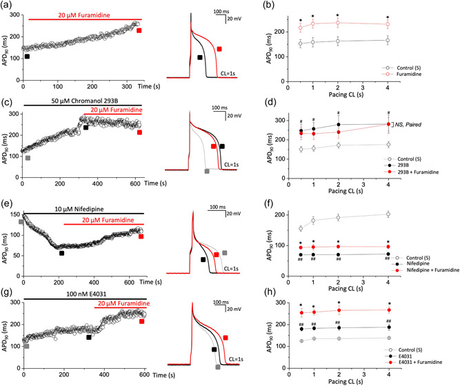
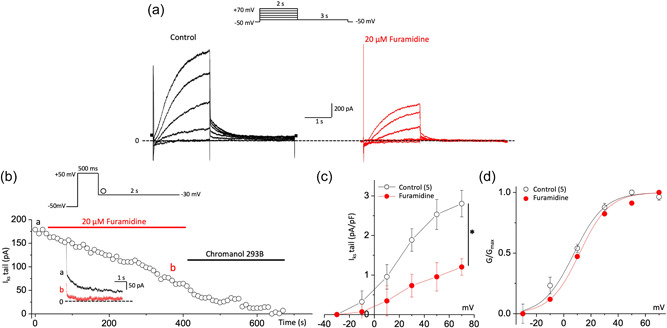
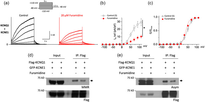
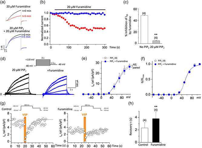
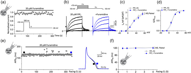
Recent studies have shown that protein arginine methyltransferase 1 (PRMT1) is highly expressed in the human heart, and loss of PRMT1 contributes to cardiac remodeling in the heart failure. However, the functional importance of PRMT1 in cardiac ion channels remains uncertain. The slow activating delayed rectifier K+ (IKs ) channel is a cardiac K+ channel composed of KCNQ1 and KCNE1 subunits and is a new therapeutic target for treating lethal arrhythmias in many cardiac pathologies, especially heart failure. Here, we demonstrate that PRMT1 is a critical regulator of the IKs channel and cardiac rhythm. In the guinea pig ventricular myocytes, treatment with furamidine, a PRMT1-specific inhibitor, prolonged the action potential duration (APD). We further show that this APD prolongation was attributable to IKs reduction. In HEK293T cells expressing human KCNQ1 and KCNE1, inhibiting PRMT1 via furamidine reduced IKs and concurrently decreased the arginine methylation of KCNQ1, a pore-forming α-subunit. Evidence presented here indicates that furamidine decreased IKs mainly by lowering the affinity of IKs channels for the membrane phospholipid, phosphatidylinositol 4,5-bisphosphate (PIP2 ), which is crucial for pore opening. Finally, applying exogenous PIP2 to cardiomyocytes prevented the furamidine-induced IKs reduction and APD prolongation. Taken together, these results indicate that PRMT1 positively regulated IKs activity through channel-PIP2 interaction, thereby restricting excessive cardiac action potential.

Keywords: PIP2; PRMT1; arrhythmia; cardiac myocytes; delayed rectifier potassium channel.


Conflict of interest statement

The authors declare no conflicts of interest.

Figures

Figure 1
Figure 1
PRMT1 inhibition with furamidine, a PRMT1 specific inhibitor, prolongs action potential duration (APD) in guinea pig ventricular myocytes via I Ks activity. (a,c,e,g) Action potentials (APs) were elicited consecutively at a pacing cycle length (CL) of 1 s and values of APD at 90% repolarization (APD90) were plotted over time. APD90 was consecutively recorded during furamidine application from a cell in control (a) or after pretreatment with chromanol 293B (c), nifedipine (e), or E4031 (g). Right, representative AP traces before (black in each panel) and after furamidine (red in each panel) treatment. AP traces before channel blocker application were also superimposed (gray in c, e, and g panels). (b,d,f,h) The summary data for the APD90 during stimulation at various pacing CLs. NS, not significantly different; *p < 0.05 control or channel blocker versus furamidine treatment; paired Student's t‐test. # p < 0.05, ## p < 0.01 control versus each channel blocker in the same group; paired Student's t‐test. All data are mean ± SEM. PRMT1, protein arginine methyltransferase 1.
Figure 2
Figure 2
PRMT1 inhibition reduces I Ks activity in guinea pig ventricular myocytes. (a) Representative current trace of I Ks before (black) and after 20 μM furamidine treatment (red) in ventricular myocytes of guinea pig. Inset shows the pulse protocol. (b) Time course of change in the amplitude of the I Ks tail (deactivation) during applications of furamidine. Representative current traces obtained at the points indicated by (a) and (b) are also shown on an expanded time scale in the inset. Currents were elicited by a voltage step to +50 mV at a holding potential of −50 mV with a subsequent step to −30 mV for the tail current. (c) The current–voltage (IV) relationships were obtained before and after the application of furamidine. Currents were elicited by voltage steps from −30 to +70 mV with a subsequent step to −30 mV for the tail current. (d) Steady‐state activation curves, with relative conductance derived from maximal chord conductance and reversal potential (E rev) for each IV, and peak I K/(E m − E rev). The resulting conductance was normalized to the maximal chord conductance. Data were fitted to a Boltzmann function (smooth curves). *p < 0.05 by paired Student's t‐test. All data are mean ± SEM. PRMT1, protein arginine methyltransferase 1.
Figure 3
Figure 3
Furamidine induces a reduction in both I Ks activity and KCNQ1 methylation in HEK 293T cells. (a) Representative current traces before (black) and after 20 μM furamidine treatment (red) from cells expressing human KCNQ1 and human KCNE1 channels. Cells were held at −80 mV, subjected to 3 s voltage steps ranging from −110 to +110 mV in 20 mV increments followed by a 1.5 s tail pulse at −40 mV (inset). (b) Tail current density–voltage relationship of I Ks measured before (black) and after exposure to furamidine (red) showed the inhibitory effect of furamidine. (c) Voltage activation curves were obtained by plotting normalized tail currents versus the prepulse potential. Values for the midpoint of activation (V 1/2) were obtained by fitting with the Boltzmann equation (lines) as described in Section 2. (d,e) HEK293T cells were transfected with an equal amount of Flag‐KCNQ1 and GFP‐KCNE1 plasmids. Immunoprecipitation was performed using anti‐Flag antibodies and monomethylation (MMR, d) or asymmetric dimethylation (ASYM, e) of the arginine residues of the precipitated proteins in the presence or absence of furamidine were analyzed by Western blotting. *p < 0.05 by paired Student's t‐test. All data are mean ± SEM. GFP, green fluorescent protein; IP, immunoprecipitation.
Figure 4
Figure 4
Furamidine decreases PIPaffinity of I Ks channels. (a,b) Furamidine‐induced inhibition of I Ks and its prevention by diC8‐PIP2 in HEK293T cells. Current traces (a) and time course (b) show furamidine‐induced inhibition of I Ks (red circles). This inhibition was prevented by the addition of 20 μM diC8‐PIP2 in the patch pipette solution (blue circles). Currents were elicited by voltage steps from −80 to +80 mV for 5 s every 10 s and normalized to currents at t = 0. (c) Histogram depicting the extent of inhibition of I Ks at +80 mV by furamidine treatment (20 μM, 5 min) in the absence or presence of 20 μM diC8‐PIP2 as indicated. **p < 0.01 by Student's t‐test. (d–f) Representative I Ks traces evoked by depolarizing voltage steps as shown in the inset in HEK293T cells loaded with 20 μM diC8‐PIP2 (d), the corresponding IV curves (e), and the corresponding voltage activation curves (f) before (black) and after 20 μM furamidine treatment (blue). NS, not significantly different; paired Student's t‐test. (g,h) Quantitative determination of the sensitivity of I Ks to activation of Dr‐VSP in HEK293T cells transfected with Dr‐VSP and human KCNQ1 + human KCNE1. Tail current amplitudes were used to measure current inhibition by Dr‐VSP activation and its recovery. Time course of I Ks tail before (left) and after 20 μM furamidine treatment (right) (g). Data were fitted with a single exponential (smooth curves). The time constant (τ) of an exponential fit of recovery before and after 20 μM furamidine treatment (h). Membrane was held at −70 mV and depolarized to +40 mV for 300 ms every 1 s, except for the shaded area in orange where the membrane was held at +100 mV for 2 s (inset). Tail currents were measured during slow channel deactivation at −30 mV. **p < 0.01 by paired Student's t‐test. Dr‐VSP, voltage‐sensitive phosphatase from Danio rerio;PIP2, phosphatidylinositol 4,5‐bisphosphate.
Figure 5
Figure 5
PIPprevented furamidine‐induced I Ks reduction and APD prolongation in cardiomyocytes. (a) The time course of I Ks tail of guinea pig ventricular myocytes shows that the inhibition I Ks by furamidine was almost completely blocked when loaded with 20 μM diC8‐PIP2. Currents were elicited by a voltage step to +50 mV at a holding potential of −50 mV with a subsequent step to −30 mV for the tail current (inset). (b–d) Representative I Ks traces evoked by depolarizing voltage steps as shown in the inset in guinea pig ventricular myocytes loaded with 20 μM diC8‐PIP2 (b), the corresponding IV curves (c), and the corresponding voltage activation curves (d) before (black) and after 20 μM furamidine treatment (blue). (e) The time course of APD90 in a guinea pig ventricular myocyte with 20 μM diC8‐PIP2 in the recording pipette in response to 20 μM furamidine. Right, representative AP trace before (black) and after (blue) 20 μM furamidine treatment from left. (f) The summary data for the APD90 from guinea pig ventricular myocytes during stimulation at various pacing CL with 20 μM diC8‐PIP2 in the patch pipette before (black) and after 20 μM furamidine treatment (blue). NS, not significantly different versus furamidine treatment; paired Student's t‐test. All data are mean ± SEM. APD, action potential duration; APD90, APD at 90% repolarization; CL, cycle length; PIP2, phosphatidylinositol 4,5‐bisphosphate.

Similar articles

Cited by

References

    1. Aflaki, M. , Qi, X. Y. , Xiao, L. , Ordog, B. , Tadevosyan, A. , Luo, X. , Maguy, A. , Shi, Y. , Tardif, J. C. , & Nattel, S. (2014). Exchange protein directly activated by cAMP mediates slow delayed‐rectifier current remodeling by sustained β‐adrenergic activation in guinea pig hearts. Circulation Research, 114(6), 993–1003. 10.1161/circresaha.113.302982 - DOI - PubMed
    1. Albrecht, L. V. , Zhang, L. , Shabanowitz, J. , Purevjav, E. , Towbin, J. A. , Hunt, D. F. , & Green, K. J. (2015). GSK3‐ and PRMT‐1‐dependent modifications of desmoplakin control desmoplakin–cytoskeleton dynamics. Journal of Cell Biology, 208(5), 597–612. 10.1083/jcb.201406020 - DOI - PMC - PubMed
    1. Bedford, M. T. , & Clarke, S. G. (2009). Protein arginine methylation in mammals: Who, what, and why. Molecular Cell, 33(1), 1–13. 10.1016/j.molcel.2008.12.013 - DOI - PMC - PubMed
    1. Bedford, M. T. , & Richard, S. (2005). Arginine methylation an emerging regulator of protein function. Molecular Cell, 18(3), 263–272. 10.1016/j.molcel.2005.04.003 - DOI - PubMed
    1. Bian, J. , Cui, J. , & McDonald, T. V. (2001). HERG K(+) channel activity is regulated by changes in phosphatidyl inositol 4,5‐bisphosphate. Circulation Research, 89(12), 1168–1176. 10.1161/hh2401.101375 - DOI - PubMed

Publication types

MeSH terms

Substances