. 2022 May 26;13(20):4563-4569.
doi: 10.1021/acs.jpclett.2c00559. Epub 2022 May 17.

Unsaturation in the Fatty Acids of Phospholipids Drastically Alters the Structure and Toxicity of Insulin Aggregates Grown in Their Presence

Affiliations

Unsaturation in the Fatty Acids of Phospholipids Drastically Alters the Structure and Toxicity of Insulin Aggregates Grown in Their Presence

Mikhail Matveyenka et al. J Phys Chem Lett. .

Abstract

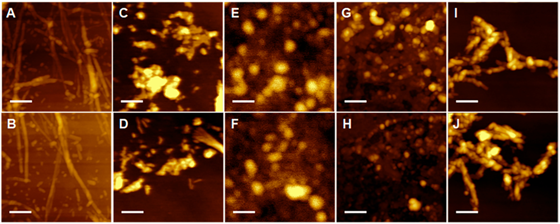
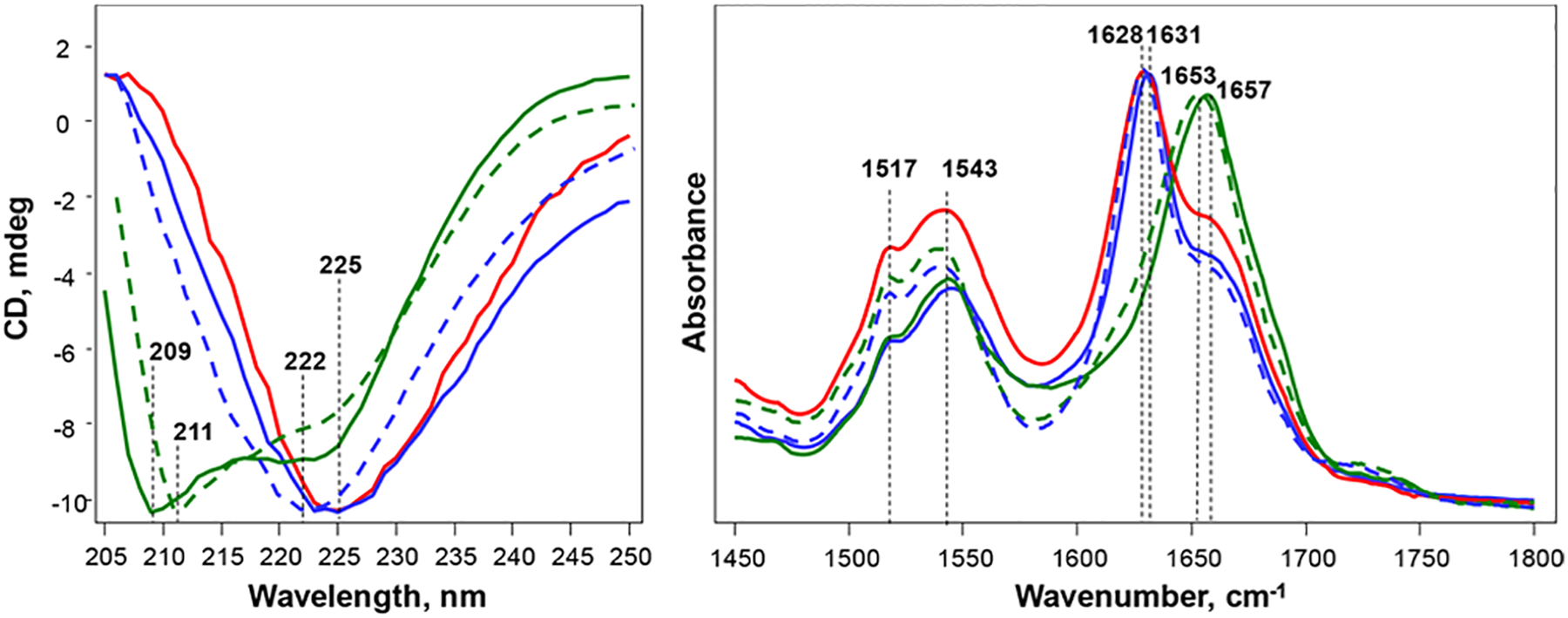
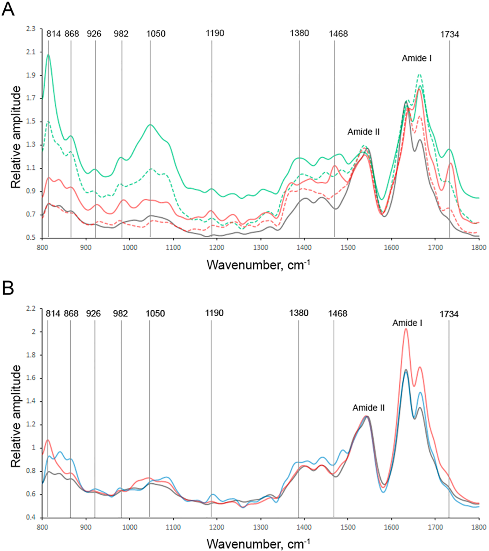
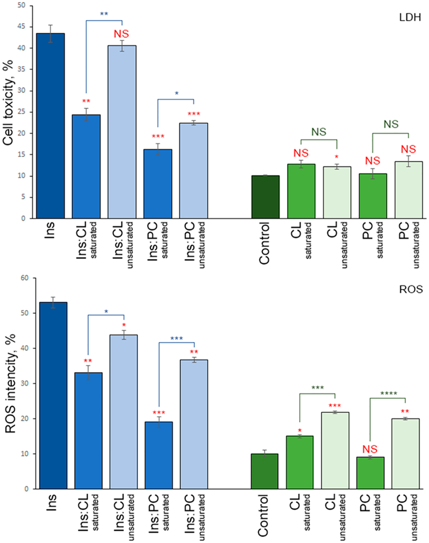
Lipid bilayers play an important role in the pathological assembly of amyloidogenic proteins and peptides. This assembly yields oligomers and fibrils, which are highly toxic protein aggregates. In this study, we investigated the role of saturation in fatty acids of two phospholipids that are present in cell membranes. We found that unsaturated cardiolipin (CL) drastically shortened the lag phase of insulin aggregation. Furthermore, structurally and morphologically different aggregates were formed in the presence of unsaturated CL vs saturated CL. These aggregates exerted drastically different cell toxicity. Both saturated and unsaturated phosphatidylcholine (PC) were able to inhibit insulin aggregation equally efficiently. Similar to CL, structurally different aggregates were formed in the presence of saturated and unsaturated PC. These aggregates exerted different cell toxicities. These results show that unsaturated phospholipids catalyze the formation of more toxic amyloid aggregates comparing to those formed in the presence of saturated lipids.


Conflict of interest statement

The authors declare no competing financial interest.

Figures

Figure 1.
Figure 1.
Lipids uniquely alter the morphologies of insulin aggregates. AFM images of (A and B) Ins:CL-u, (C and D) Ins:CL-s, (E and F) Ins:PC-u, (G and H) Ins:PC-s, and (I and J) insulin aggregates grown in the lipid-free environment. Scale bars are 200 nm.
Figure 2.
Figure 2.
Structural analysis of insulin aggregates. (Left) CD and (right) ATR-FTIR spectra of insulin aggregates (Ins) grown in the lipid-free environment (red) as well as in the presence of Ins:CL-s (solid blue), Ins:CL-u (dashed blue), Ins:PC-s (solid green), and Ins:PC-u (dashed green).
Figure 3.
Figure 3.
Nanoscale analysis of (A) Ins:PC-u (red) and Ins:PC-s (green) and (B) Ins:CL-u (black) and Ins:CL-s (blue) aggregates. Spectra collected from individual aggregates are shown in the SI. AFM–IR spectra of population A are shown in the corresponding solid, whereas population B is shown by the corresponding dashed lines.
Figure 4.
Figure 4.
Insulin aggregates grown in the presence of saturated lipids possess cell toxicity different from that of the aggregates grown in the presence of unsaturated lipids. Histograms of (top) LDH and (bottom) ROS assays of Ins, Ins:CL-s, Ins:CL-u, Ins:PC-s, and Ins:PC-u (blue bars) and saturated and unsaturated lipids (green bars) themselves. For LDH and ROS production, error bars represent the means of three replicates. Red asterisks (*) show the significance of the level of difference between Ins and Ins aggregates grown in the presence of lipids as well as between lipid samples and the control. Blue asterisks show the significance of the level of difference between protein samples with saturated lipids and unsaturated lipids. NS is a nonsignificant difference, and *p ≤ 0.05, **p ≤ 0.01, and ***p ≤ 0.001.

Similar articles

Cited by

References

    1. van Meer G; Voelker DR; Feigenson GW Membrane Lipids: Where They Are and How They Behave. Nat. Rev. Mol. Cell Biol 2008, 9, 112–24. - PMC - PubMed
    1. Fahy E; Subramaniam S; Murphy RC; Nishijima M; Raetz CR; Shimizu T; Spener F; van Meer G; Wakelam MJ; Dennis EA Update of the Lipid Maps Comprehensive Classification System for Lipids. J. Lipid Res 2009, 50 (Suppl), S9–14. - PMC - PubMed
    1. Fitzner D; Bader JM; Penkert H; Bergner CG; Su M; Weil MT; Surma MA; Mann M; Klose C; Simons M Cell-Type- and Brain-Region-Resolved Mouse Brain Lipidome. Cell Rep 2020, 32, 108132. - PubMed
    1. Pope S; Land JM; Heales SJ Oxidative Stress and Mitochondrial Dysfunction in Neurodegeneration; Cardiolipin a Critical Target? Biochim. Biophys. Acta 2008, 1777, 794–9. - PubMed
    1. Falabella M; Vernon HJ; Hanna MG; Claypool SM; Pitceathly RDS Cardiolipin, Mitochondria, and Neurological Disease. Trends Endocrinol. Metab 2021, 32, 224–237. - PMC - PubMed