. 2022 Jun 13;8(6):2537-2552.
doi: 10.1021/acsbiomaterials.2c00392. Epub 2022 May 17.

Role of Nitric Oxide-Releasing Glycosaminoglycans in Wound Healing

Affiliations

Role of Nitric Oxide-Releasing Glycosaminoglycans in Wound Healing

Sara E Maloney et al. ACS Biomater Sci Eng. .

Abstract

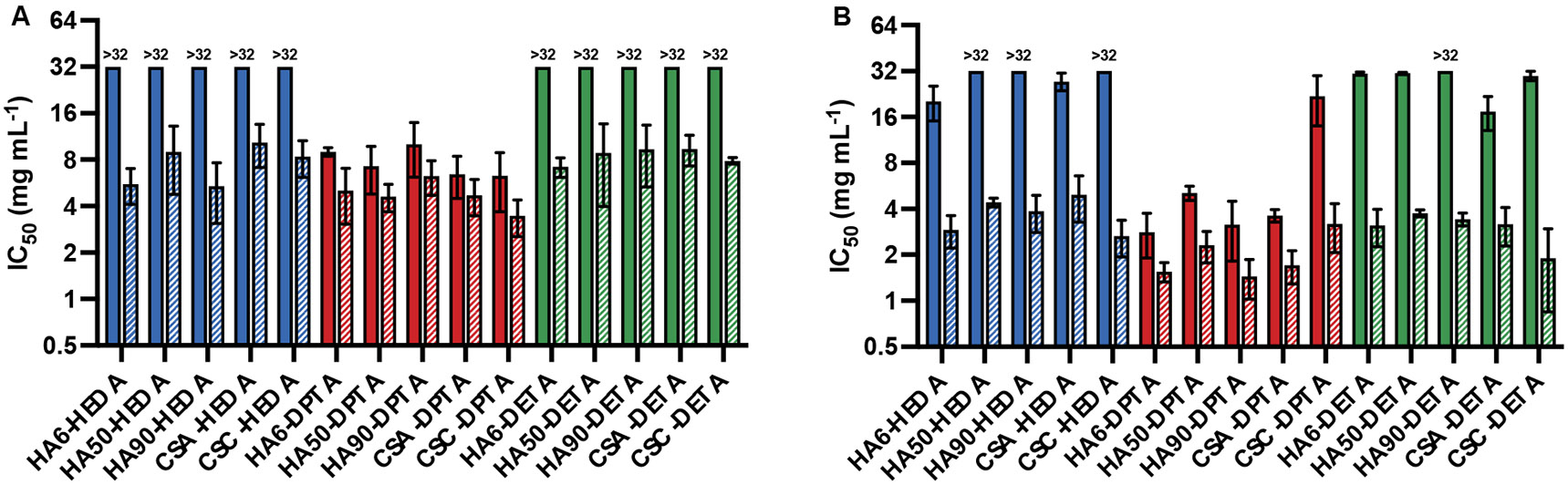
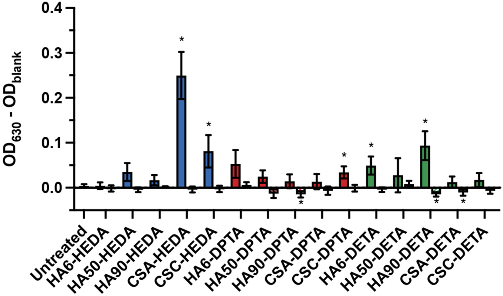
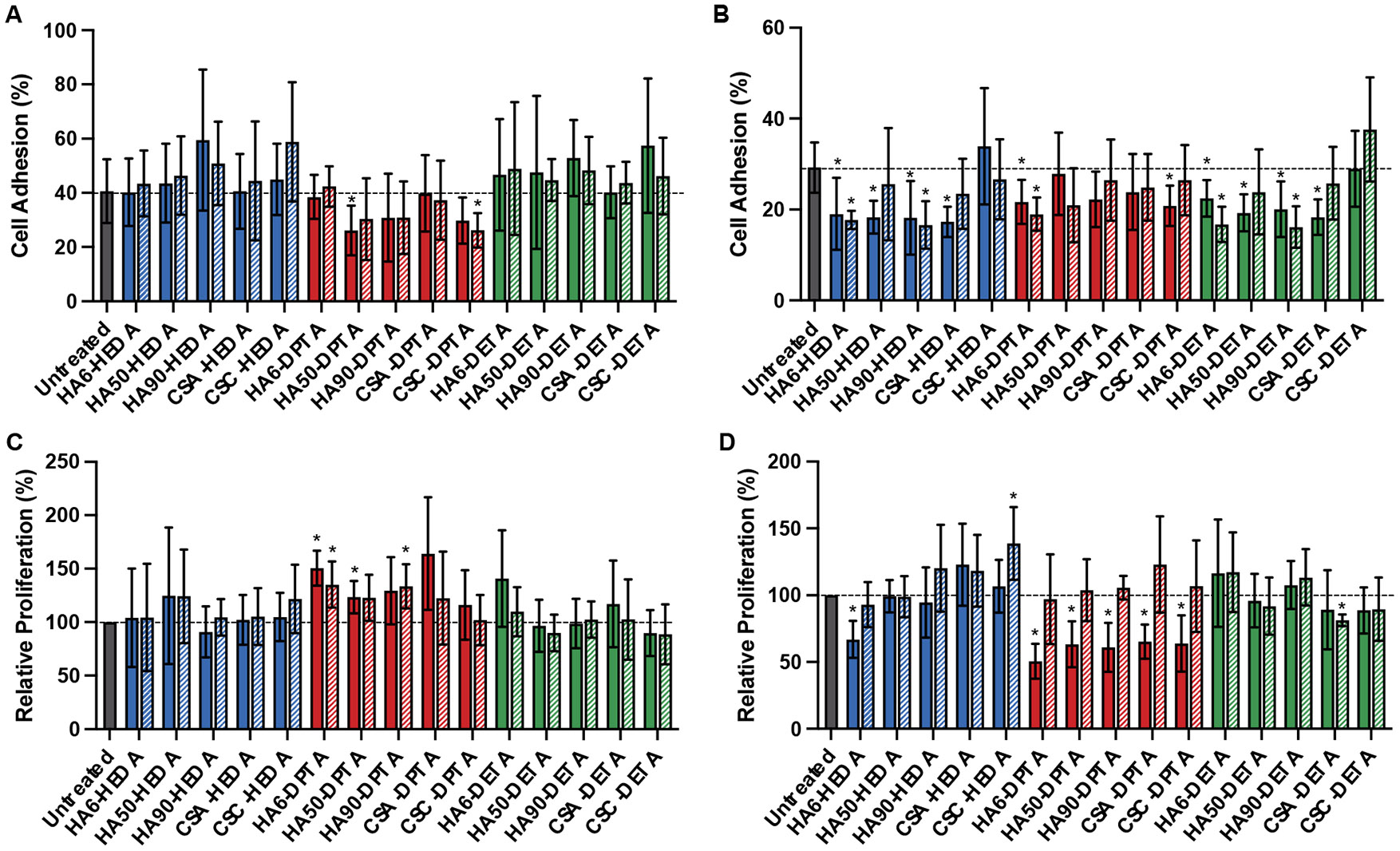
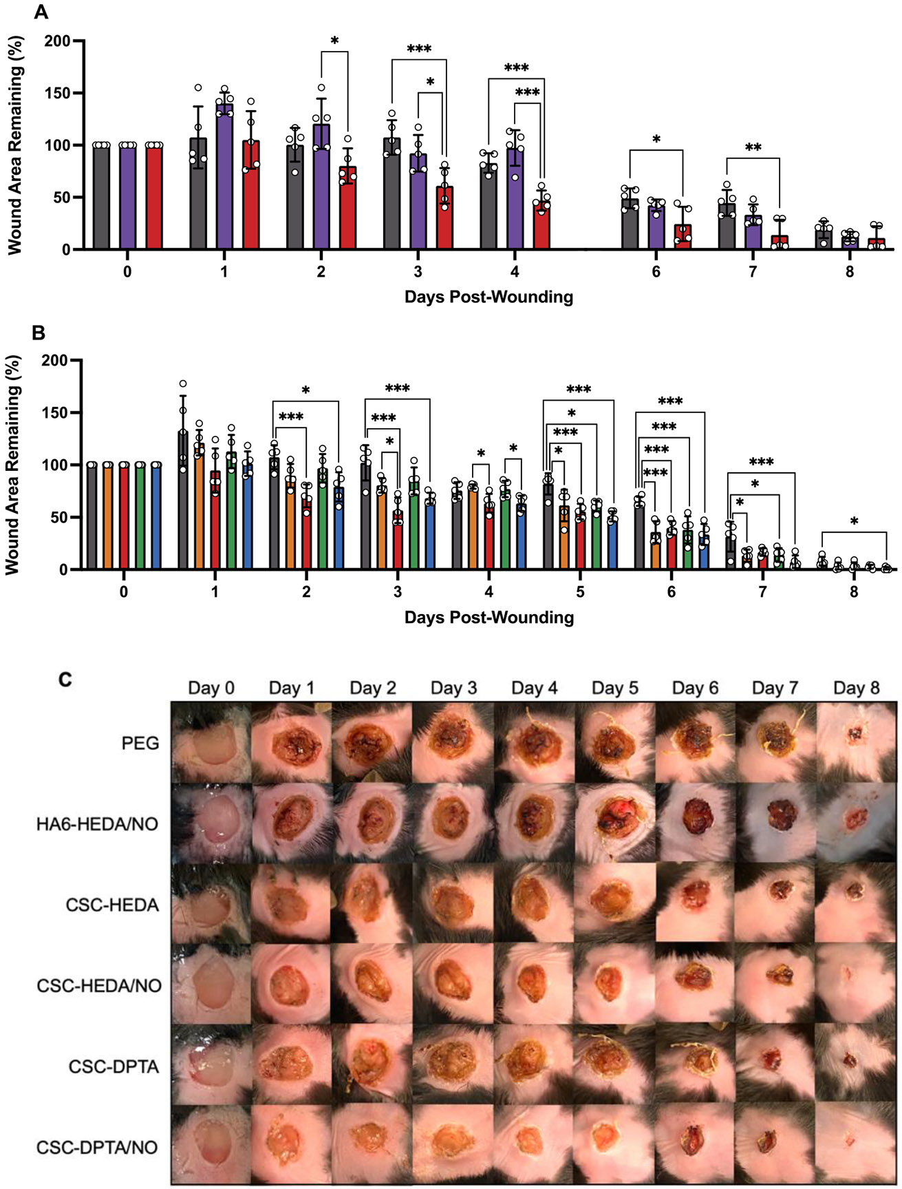
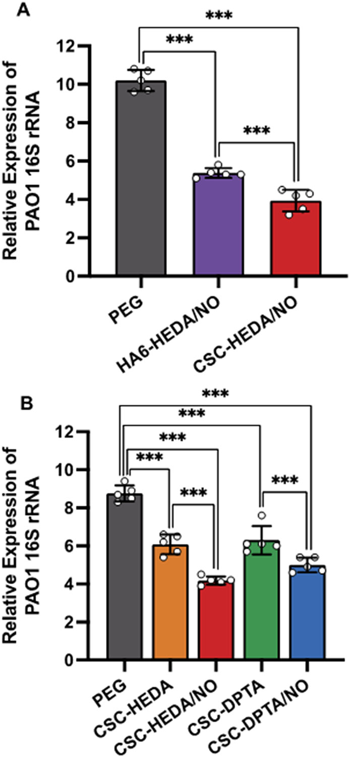
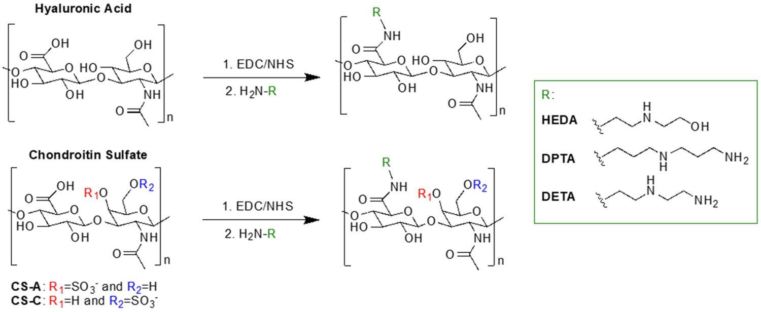
Two glycosaminoglycan (GAG) biopolymers, hyaluronic acid (HA) and chondroitin sulfate (CS), were chemically modified via carbodiimide chemistry to facilitate the loading and release of nitric oxide (NO) to develop a multi-action wound healing agent. The resulting NO-releasing GAGs released 0.2-0.9 μmol NO mg-1 GAG into simulated wound fluid with NO-release half-lives ranging from 20 to 110 min. GAGs containing alkylamines with terminal primary amines and displaying intermediate NO-release kinetics exhibited potent, broad spectrum bactericidal action against three strains each of Pseudomonas aeruginosa and Staphylococcus aureus ranging in antibiotic resistance profile. NO loading of the GAGs was also found to decrease murine TLR4 activation, suggesting that the therapeutic exhibits anti-inflammatory mechanisms. In vitro adhesion and proliferation assays utilizing human dermal fibroblasts and human epidermal keratinocytes displayed differences as a function of the GAG backbone, alkylamine identity, and NO-release properties. In combination with antibacterial properties, the adhesion and proliferation profiles of the GAG derivatives enabled the selection of the most promising wound healing candidates for subsequent in vivo studies. A P. aeruginosa-infected murine wound model revealed the benefits of CS over HA as a pro-wound healing NO donor scaffold, with benefits of accelerated wound closure and decreased bacterial burden attributable to both active NO release and the biopolymer backbone.

Keywords: antibacterial; chondroitin sulfate; hyaluronic acid; nitric oxide; wound healing.


Figures

Figure 1.
Figure 1.
Concentration of amine-modified (solid) or NO-releasing (striped) glycosaminoglycan derivatives required to inhibit metabolic activity of (A) human dermal fibroblasts (HDFs) or (B) human epidermal keratinocytes (HEKs) by 50% (IC50).
Figure 2.
Figure 2.
Activation of NF-κB via murine TLR4 receptor in HEK-Blue mTLR4 cells upon treatment with amine-modified (solid) or NO-releasing (striped) GAG derivatives at a concentration of 100 μg mL−1. NF-κB-induced SEAP activity is reported as the OD630 corrected for blank media. Error bars represent the standard deviation of n ≥ 3 separate experiments. * p < 0.05 compared to untreated cells.
Figure 3.
Figure 3.
(A, B) Adhesion and (C, D) relative proliferation of (A, C) HDFs and (B, D) HEKs treated with amine-modified (solid) or NO-releasing (striped) GAG derivatives at a concentration of 10 μg mL−1. Adhesion of GAG derivatives is reported as a percentage of the total number of cells seeded in each well. Proliferation of GAG derivatives is reported relative to untreated cells (set to 100% proliferation). Error bars represent the standard deviation of n ≥ 4 separate experiments. * p < 0.05 compared to untreated cells. Note: the y-axis is scaled appropriately for each data set.
Figure 4.
Figure 4.
(A) Percentage of initial wound area remaining following daily treatment with PEG (gray), 50 mg kg−1 of HA6-HEDA/NO in PEG (purple), or 50 mg kg−1 of CSC-HEDA/NO in PEG (red). Of note, mice were treated and imaged on day 5 post-wounding but not measured. (B) Percentage of initial wound area remaining following daily treatment with PEG (gray), 50 mg kg−1 of CSC-HEDA in PEG (orange), 50 mg kg−1 of CSC-HEDA/NO in PEG (red), 50 mg kg−1 of CSC-DPTA in PEG (green), or 50 mg kg−1 of CSC-DPTA/NO in PEG (blue). (C) Representative images of wounds from each treatment group. Error bars represent the standard deviation for n = 5 mice. *p < 0.05, **p < 0.01, ***p < 0.005.
Figure 5.
Figure 5.
Relative quantity of P. aeruginosa genome remaining in wound tissue harvested 8 days post-wounding for mice treated daily with (A) PEG (gray), 50 mg kg−1 of HA6-HEDA/NO in PEG (purple), or 50 mg kg−1 of CSC-HEDA/NO in PEG (red), or (B) PEG (gray), 50 mg kg−1 of CSC-HEDA in PEG (orange), 50 mg kg−1 of CSC-HEDA/NO in PEG (red), 50 mg kg−1 of CSC-DPTA in PEG (green), or 50 mg kg−1 of CSC-DPTA/NO in PEG (blue). Error bars represent the standard deviation for n=5 mice. *p < 0.05, ** p < 0.01, *** p < 0.005.
Scheme 1.
Scheme 1.
Modification of hyaluronic acid and chondroitin sulfate with alkylamines.

Similar articles

Cited by

References

    1. Sen CK; Gordillo GM; Roy S; Kirsner R; Lambert L; Hunt TK; Gottrup F; Gurtner GC; Longaker MT Human Skin Wounds: A Major and Snowballing Threat to Public Health and the Economy. Wound Repair Regen. 2009, 17, 763–771. - PMC - PubMed
    1. Sen CK Human Wounds and Its Burden: An Updated Compendium of Estimates. Adv. Wound Care 2019, 8, 39–48. - PMC - PubMed
    1. Järbrink K; Ni G; Sönnergren H; Schmidtchen A; Pang C; Bajpai R; Car J The Humanistic and Economic Burden of Chronic Wounds: A Protocol for a Systematic Review. Syst. Rev 2017, 6, 1–7. - PMC - PubMed
    1. Han G; Ceilley R Chronic Wound Healing: A Review of Current Management and Treatments. Adv. Ther 2017, 34, 599–610. - PMC - PubMed
    1. Stechmiller JK; Childress B; Cowan L Arginine Supplementation and Wound Healing. 2005, 52–61. - PubMed

Publication types

LinkOut - more resources