. 2022 May;10(5):e004007.
doi: 10.1136/jitc-2021-004007.

Antigen mimicry as an effective strategy to induce CSPG4-targeted immunity in dogs with oral melanoma: a veterinary trial

Affiliations

Antigen mimicry as an effective strategy to induce CSPG4-targeted immunity in dogs with oral melanoma: a veterinary trial

Federica Riccardo et al. J Immunother Cancer. 2022 May.

Abstract

Background: Melanoma is the most lethal form of skin cancer in humans. Conventional therapies have limited efficacy, and overall response is still unsatisfactory considering that immune checkpoint inhibitors induce lasting clinical responses only in a low percentage of patients. This has prompted us to develop a vaccination strategy employing the tumor antigen chondroitin sulfate proteoglycan (CSPG)4 as a target.

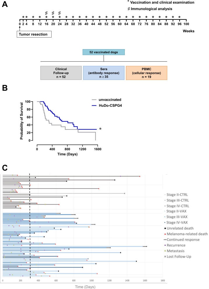
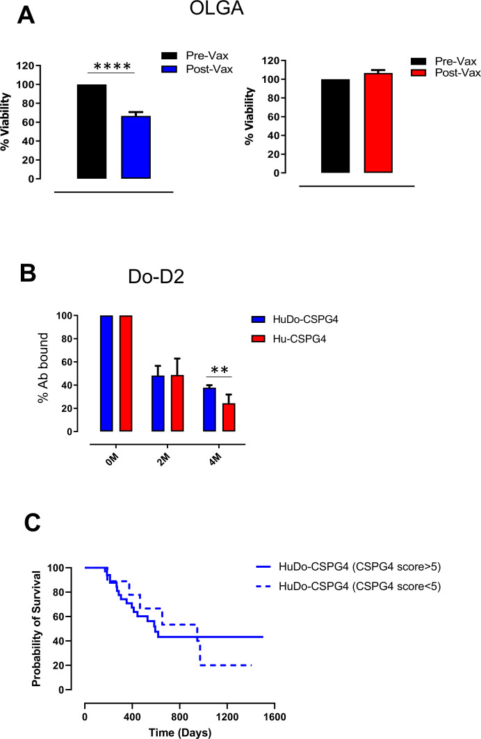
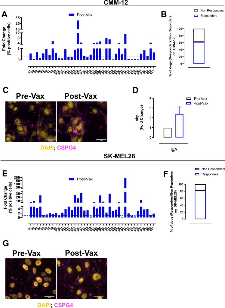
Methods: To overcome the host's unresponsiveness to the self-antigen CSPG4, we have taken advantage of the conservation of CSPG4 sequence through phylogenetic evolution, so we have used a vaccine, based on a chimeric DNA molecule encompassing both human (Hu) and dog (Do) portions of CSPG4 (HuDo-CSPG4). We have tested its safety and immunogenicity (primary objectives), along with its therapeutic efficacy (secondary outcome), in a prospective, non-randomized, veterinary clinical trial enrolling 80 client-owned dogs with surgically resected, CSPG4-positive, stage II-IV oral melanoma.

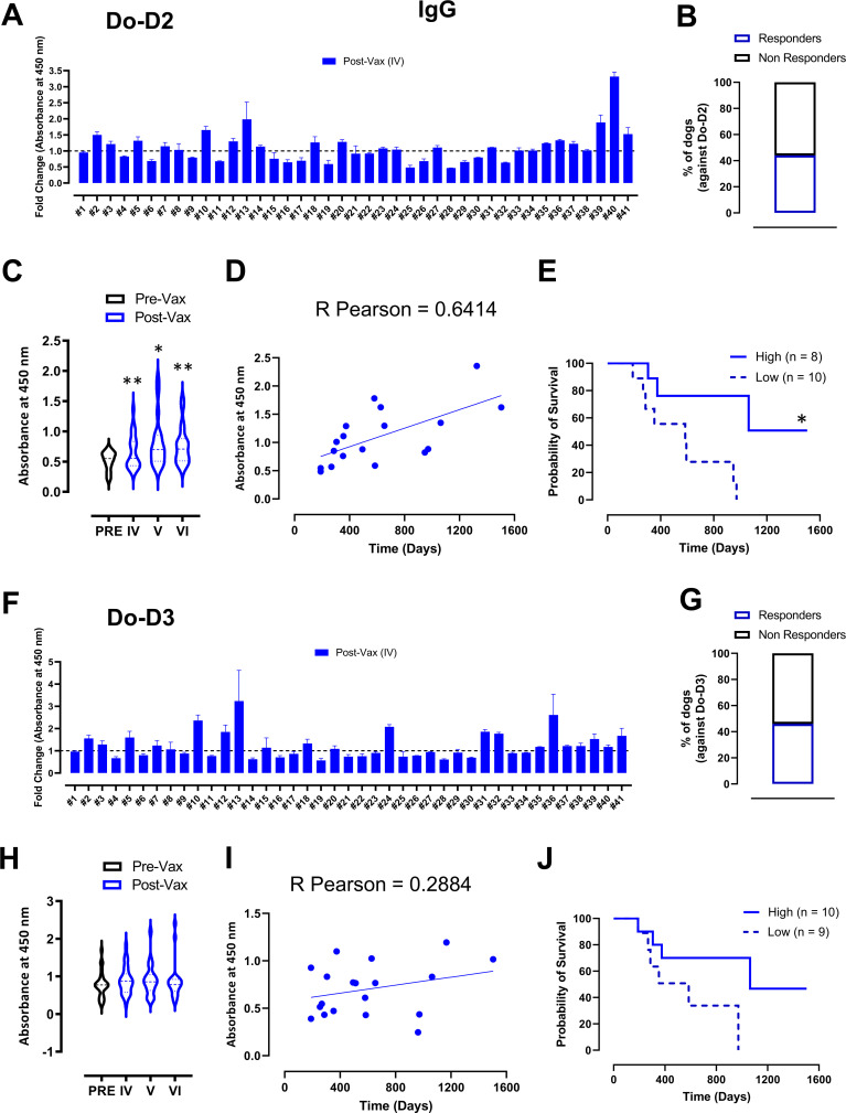
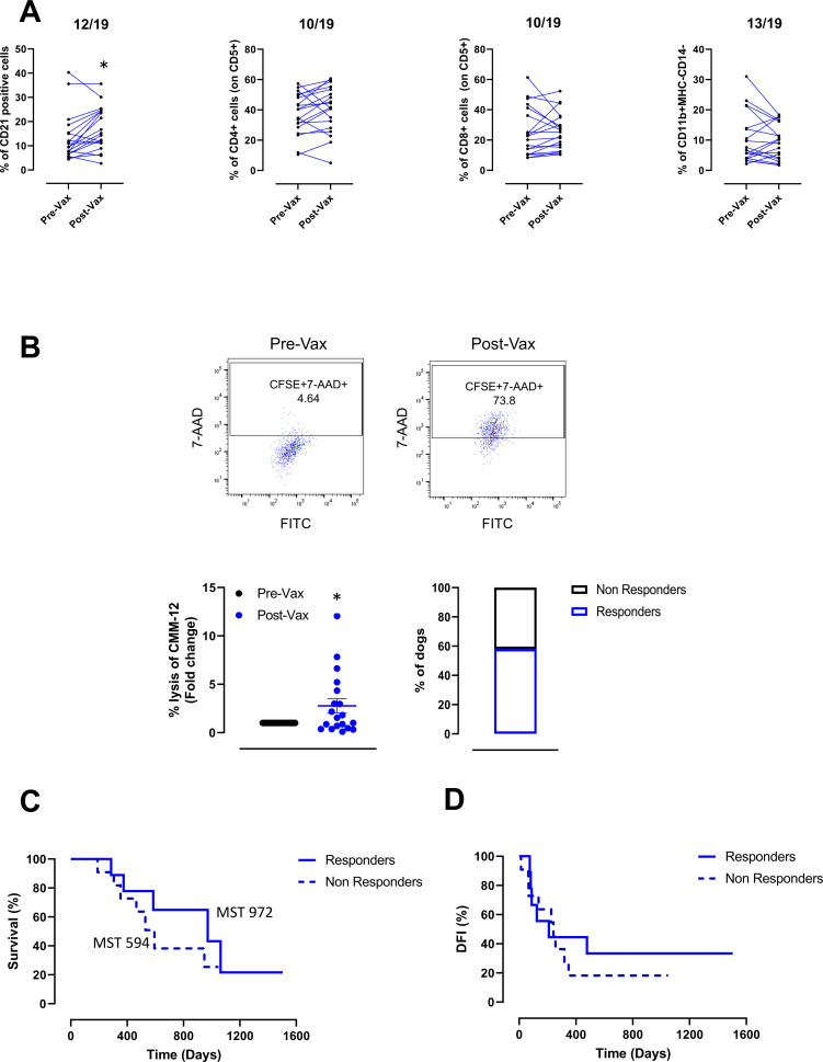
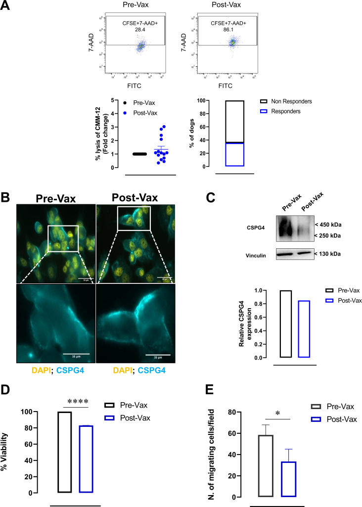
Results: Vaccinated dogs developed anti-Do-CSPG4 and Hu-CSPG4 immune response. Interestingly, the antibody titer in vaccinated dogs was significantly associated with the overall survival. Our data suggest that there may be a contribution of the HuDo-CSPG4 vaccination to the improvement of survival of vaccinated dogs as compared with controls treated with conventional therapies alone.

Conclusions: HuDo-CSPG4 adjuvant vaccination was safe and immunogenic in dogs with oral melanoma, with potential beneficial effects on the course of the disease. Thanks to the power of naturally occurring canine tumors as predictive models for cancer immunotherapy response, these data may represent a basis for the translation of this approach to the treatment of human patients with CSPG4-positive melanoma subtypes.

Keywords: Immunogenicity, Vaccine; Immunotherapy, Active; Melanoma; Translational Medical Research; Vaccination.


Conflict of interest statement

Competing interests: No, there are no competing interests.

Figures

Figure 1
Figure 1
Adjuvant chimeric HuDo-CSPG4 vaccination improves the survival of canine patients affected by CSPG4-positive oral melanomas. (A) Immunization protocol (upper panel) and study design (lower panel). (B) Kaplan-Meier curves comparing the overall survival (in days) of HuDo-CSPG4 vaccinated (blue line) and unvaccinated (gray line) dogs, after the local control of CSPG4-positive oral melanoma, updated to December 2021. Log-rank test, *p=0.0320. (C) Swimmer plot depicting the overall survival of canine melanoma patients enrolled in the study. Briefly, the survival (in days) of dogs with surgically resected CSPG4-positive melanoma, either vaccinated (Vax) or non-vaccinated (Ctrl), is reported, considering the day 0 as the moment of the surgery for each dog. Arrows indicate that the patients were still alive at the time of publication (continued response). For each patient, the moment of recurrence or metastasis detection, if any has been indicated. Black dots indicate patients who died because of unrelated reasons, while red triangles indicate patients who died because of melanoma. Dogs lost in the follow-up (n=3) were also indicated. The median survival time (310 days) for the control group has been indicated by a dotted vertical line. CSPG4, chondroitin sulfate proteoglycan 4; PBMC, peripheral blood mononuclear cells.
Figure 2
Figure 2
HuDo-CSPG4 vaccination is effective in inducing a specific anti-canine CSPG4 antibody response in dogs. (A) and (F) Analysis of the presence of IgG antibodies against the Do–D2 (A) and Do–D3 (F) domains of the canine CSPG4 protein in the sera of dogs before the first immunization (Pre-Vax, dotted black line) and after the fourth HuDo-CSPG4 vaccination (Post-Vax, blue bars), measured by ELISA. Results are expressed as the ratio (fold change) between the absorbance measured at 450 nm of the Post-Vax and the Pre-Vax sera. Each bar represents a canine patient. (B) and (G) Histograms representing the percentage of responders (blue) and non-responders (black) calculated by enumerating dogs in which sera collected after the fourth immunization displayed an increased ability to bind the Do–D2 (B) and the Do–D3 (G) domains as compared with sera collected before the first immunization. (C) and (H) Violin graphs representing the absorbance measured at 450 nm by ELISA against the Do–D2 (C) and Do–D3 (H) of pre-vaccination and post-vaccination sera from dogs who respond after the fourth, the fifth or the sixth immunizations. Student’s t-test, p*=0–0108, p**<0.0085. (D) and (I) Correlation between the absorbance values measured at 450 nm by ELISA of the post-vaccination IgG of responder dogs against the Do–D2 (D) and Do–D3 (I) domains and the overall survival. Each dot represents a responder dog. Pearson correlation coefficients (r) are shown. (E) and (J) Kaplan-Meier curves correlating the overall survival of vaccinated dogs who develop (responders) a specific IgG response against the Do–D2 (E) and Do–D3 (J) domains with a high (continuous blue lines) or low (dotted blue lines) antibody level measured in their post-vaccination sera by ELISA, considering as cut-off the mean of the absorbance at 450 nm. Log-rank test, *p=0.0120. CSPG4, chondroitin sulfate proteoglycan 4; Do, dog; Hu, human.
Figure 4
Figure 4
HuDo-CSPG4 vaccination is effective in inducing an anti-CSPG4 cellular immune response in dogs. (A) Flow cytometry analysis of the frequency of circulating B cells, CD4+ and CD8+ T cells, and MDSC collected from canine melanoma patients before (Pre-Vax) and after the fourth HuDo-CSPG4 vaccination (Post-Vax). Graphs show the percentage of CD21+ B cells (gated on live cells), of CD4+ and CD8+ T cells (gated on CD5+ cells) and of MHC-IICD14 (gated on CD11b+ cells) cells. The numbers of dogs in which a difference in the frequency (fold change >1.1 or fold change <1.1) of a cell population was observed comparing Pre-Vax and Post-Vax PBMC are indicated above in each graph. Student’s t-test, *p=0.0151. (B) Cytotoxic assays to quantify the ability of Pre-Vax and Post-Vax PBMC to kill CSPG4-positive CMM-12 cells. Representative dot plots of one dog analyzed, showing the percentage of 7-AAD+ dead cells among CFSE+ cells (upper panels) are shown. Results are shown as the fold change between the percentage of CMM-12 cells lysed after incubation with Post-Vax and Pre-Vax PBMC for each dog analyzed (lower, left panel, Student’s t-test, *p=0.0260), and as the percentage of dogs of which PBMC induced an increased CMM-12 cell lysis (responders) or not (non responders) (lower, right panel). (C) and (D) Kaplan-Meier curves comparing the overall survival (C) and the disease-free-interval (DFI, (D), in days, of vaccinated dogs who develop (responders, continuous blue line) or not (non responders, dotted blue line) a cytotoxic response against the canine CMM-12 cell line. The median survival times (MST) in days for each group has been reported in the overall survival graph. Log-rank test, p=0.2819. 7-AAD, 7-Amino-ActinomycinD; CSPG4, chondroitin sulfate proteoglycan 4; Do, dog; Hu, human; MDSC, myeloid derived suppressor cells; PBMC, peripheral blood mononuclear cells; FITC, fluorescein isothiocyanate; MHC, major histocompatibility complex; CFSE, carboxyfluorescein succinimidyl ester.
Figure 5
Figure 5
Potential mechanisms of action of HuDo-CSPG4 vaccine-induced antibodies. (A) Cytotoxic assay to quantify the ability of Pre-Vax and Post-Vax sera to induce the killing (ADCC) of CSPG4-positive CMM-12 cells. Representative dot plots of one dog analyzed, showing the percentage of 7-AAD+ dead cells among CFSE+ cells (upper panels) are shown. Results are reported as the fold change between the percentage of CMM-12 cells lysed after incubation with Post-Vax and Pre-Vax sera for each dog analyzed (lower, left panel), and the percentage of dogs whose sera induced an increased CMM-12 cell lysis (responders) or not (non responders) (lower right panel). (B) Representative immunofluorescence images (one out of three independent experiments) of canine CMM-12 cells incubated at 37°C with pooled sera, collected before (Pre-Vax) and after the fourth HuDo-CSPG4 vaccination (Post-Vax). Anti-CSPG4 IgG binding and localization was detected using a Texas red-conjugated anti-mouse IgG and nuclei were stained with DAPI. (C) Representative Western blot analyses (upper panel) of CSPG4 expression in the lysates of CMM-12 melanoma cells incubated at 37°C for 48 hours with pooled sera collected before (Pre-Vax) and after the fourth HuDo-CSPG4 vaccination (Post-Vax). Relative protein loading was shown using an anti-vinculin antibody. Immunoreactive band density quantification is shown (lower panel); results are reported as relative CSPG4 protein expression, considering Pre-Vax condition as 1. (D) MTT proliferation assay performed on CSPG4-positive canine CMM-12 cells after 48 hours of incubation at 37°C with pool of canine sera collected before (Pre-Vax) or after the fourth HuDo-CSPG4 vaccination (Post-Vax). Results are expressed as the percentage (%) of cell viability, considering Pre-Vax conditions as 100%. Student’s t-test, ****p<0.0001. (E) Migratory ability of canine CMM-12 melanoma cells incubated with pool of canine sera collected before (Pre-Vax) or after the fourth HuDo-CSPG4 vaccination (Post-Vax). Results show the number of migrated cells in four randomly selected fields per well. Student’s t-test, *p=0.0155. 7-AAD, 7-Amino-ActinomycinD; ADCC, antibody-dependent cell-mediated cytotoxicity; CSPG4, chondroitin sulfate proteoglycan 4; DAPI, diamidino-2-phenylindole; Do, dog; Hu, human; FITC, fluorescein isothiocyanate; MTT, 3-(4, 5-dimethylthiazolyl-2)-2,5-diphenyltetrazolium bromide; CFSE, carboxyfluorescein succinimidyl ester.
Figure 6
Figure 6
HuDo-CSPG4 vaccination induces a high avidity antibody response. (A) MTT proliferation assay performed on a low CSPG4-expressing canine melanoma cell line (OLGA) incubated for 72 hours at 37°C with pool of canine sera collected before (Pre-Vax) or after the fourth HuDo-CSPG4 (blue bars) or Hu-CSPG4 (red bars) vaccination. Results are expressed as percentage (%) of viability, considering Pre-Vax conditions as 100%. Student’s t-test, ****p=0.0002. (B) Avidity of anti-Do–D2 vaccine-induced antibodies in the sera of dogs immunized with either the HuDo-CSPG4 (blue bars) or the (Hu)-CSPG4 (red bars) plasmids, evaluated by a chaotropic ELISA. Results are expressed as percentage (%) of antibodies (Ab) that remain bound after the treatment with the chaotropic agent, as compared with the medium alone considered as 100%. Student’s t-test, **p=0.0050. (C) Kaplan-Meier curves comparing overall survival of HuDo-CSPG4 vaccinated dogs bearing a melanoma with CSPG4-positivity score <5 (dotted blue line) and ≥5 (continuous blue line). CSPG4, chondroitin sulfate proteoglycan 4; Do, dog; Hu, human; MTT, 3-(4, 5-dimethylthiazolyl-2)-2,5-diphenyltetrazolium bromide).
Figure 3
Figure 3
HuDo-CSPG4 vaccine-induced antibodies bind CSPG4-overexpressing canine and human melanoma cells. (A) and (E) Flow cytometry analysis of naturally CSPG4-expressing canine CMM-12 (A) and human SK-MEL-28 (E) melanoma cells incubated with sera from canine patients before the first immunization (Pre-Vax, dotted black line) and after the fourth HuDo-CSPG4 vaccination (Post-Vax, blue bars). Total IgG binding was evaluated using a FITC-conjugated goat anti-dog IgG secondary antibody. Results are expressed as the ratio (fold change) between the percentages (%) of stained cells incubated with the Post-Vax and the Pre-Vax sera. Each bar represents a canine patient. (B) and (F) Histograms representing the percentage of responders (blue) and non-responders (black) calculated by enumerating dogs in which sera collected after the fourth immunization displayed an increased ability (ratio >1.1) to stain the CMM-12 (B) and the SK-MEL-28 (F) melanoma cells as compared with sera collected before the first immunization. (C) and (G) Representative immunofluorescence images (one out of three independent experiments) of canine CMM-12 (C) and human SK-MEL-28 (G) cells stained with Pre-Vax and Post-Vax sera from HuDo-CSPG4 vaccinated dogs. Bound antibodies were revealed using a FITC rabbit anti-dog IgG secondary antibody and nuclei were stained with DAPI. (D) IgA specific binding of Pre-Vax and Post-Vax sera collected from HuDo-CSPG4 vaccinated dogs on canine CMM-12 cells. Results are expressed as the ratio (fold change) between the serum binding potential (sbp) of the Post-Vax and the Pre-Vax sera. CSPG4, chondroitin sulfate proteoglycan 4; DAPI, diamidino-2-phenylindole; Do, dog; Hu, human; FITC, fluorescein isothiocyanate.

Similar articles

Cited by

References

    1. Siegel RL, Miller KD, Jemal A. Cancer statistics, 2020. CA Cancer J Clin 2020;70:7–30. 10.3322/caac.21590 - DOI - PubMed
    1. Bastian BC. The molecular pathology of melanoma: an integrated taxonomy of melanocytic neoplasia. Annu Rev Pathol 2014;9:239–71. 10.1146/annurev-pathol-012513-104658 - DOI - PMC - PubMed
    1. Shaughnessy M, Klebanov N, Tsao H. Clinical and therapeutic implications of melanoma genomics. JTGG 2018;2. 10.20517/jtgg.2018.25 - DOI
    1. Petrova V, Arkhypov I, Weber R. Modern aspects of immunotherapy with checkpoint inhibitors in melanoma. Int. J. Mol. Sci 2020. 10.3390/ijms21072367 - DOI - PMC - PubMed
    1. Kuryk L, Bertinato L, Staniszewska M, et al. . From conventional therapies to immunotherapy: melanoma treatment in review. Cancers 2020;12:3057–20. 10.3390/cancers12103057 - DOI - PMC - PubMed

Publication types