Case Reports
. 2022 May 17;15(5):e248181.
doi: 10.1136/bcr-2021-248181.

Rare paediatric case of agenesis of the vermiform appendix, ileal duplication and sickle cell disease

Affiliations
Case Reports

Rare paediatric case of agenesis of the vermiform appendix, ileal duplication and sickle cell disease

Nadia Laezza et al. BMJ Case Rep. .

Abstract

This study reports an exceptional case of a 14-year-old girl with sickle cell disease that was diagnosed with agenesis of the vermiform appendix and ileal duplication. Both consist of extremely rare gastrointestinal malformations whose association has never been described. The preadolescent girl presented with abdominal pain and vomiting, and the ultrasound was suggestive of acute appendicitis. Surgical findings were agenesis of the vermiform appendix and a T-shaped ileal malformation with inflammatory changes. The patient underwent resection and ileal end-to-end anastomosis. Histopathological evaluation identified an ileal duplication, with small bowel and colonic mucosa, no communication to the adjacent ileum and ischaemic changes. At 8-month follow-up, the patient was asymptomatic.

Keywords: Congenital disorders; Gastrointestinal surgery; Haematology (incl blood transfusion); Paediatric Surgery.


Conflict of interest statement

Competing interests: None declared.

Figures

Figure 1
Figure 1
Classification of the agenesis of the vermiform appendix. Type I - absence of appendix and cecum; type II - rudimentary cecum and absence of appendix; type III - normal cecum and absence of appendix; type IV - normal cecum and rudimentary appendix; type V - giant cecum and absence of appendix. Illustrator: Vieira EPL, corresponding author of the article ‘Congenital abnormalities and anatomical variations of the vermiform appendix and mesoappendix’, and which authorised the use of the figure.
Figure 2
Figure 2
Ultrasound, showing hyperechoic fat and a stratified tubular structure (arrow), suggestive of acute appendicitis.
Figure 3
Figure 3
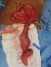
Terminal ileum (*distal segment) and ileal duplication (large arrow): a tubular structure at 10 cm from the ileocecal valve, arising from the antimesenteric border (white arrow — proximal end of the duplication), with a separate mesenteric pedicle (blue arrow), blind-ended, with 15 cm of length and marked inflammatory changes, without perforation.
Figure 4
Figure 4
Compiled image showing normal ileal segment and adjacent intestinal duplication, with a common wall, H&E.
Figure 5
Figure 5
Histological section showing two ileal type mucosal layers sharing a muscular layer, H&E x40.
Figure 6
Figure 6
Distal area with colonic mucosa with ‘pseudomembranous-like’ ischaemic changes, H&E x40.

Similar articles

References

    1. Vaos G, Misiakos EP. Congenital anomalies of the gastrointestinal tract diagnosed in adulthood--diagnosis and management. J Gastrointest Surg 2010;14:916–25. 10.1007/s11605-009-1124-z - DOI - PubMed
    1. Ramakrishna HK. Intestinal duplication. Indian J Surg 2008;70:270–3. 10.1007/s12262-008-0082-0 - DOI - PMC - PubMed
    1. Russo G, De Franceschi L, Colombatti R, et al. . Current challenges in the management of patients with sickle cell disease - A report of the Italian experience. Orphanet J Rare Dis 2019;14:120. 10.1186/s13023-019-1099-0 - DOI - PMC - PubMed
    1. Collins DC. A study of 50,000 specimens of the human vermiform appendix. Surg Gynecol Obstet 1955;101:437–45. - PubMed
    1. Vieira EPL, Bonato LM, Silva GGPda, et al. . Congenital abnormalities and anatomical variations of the vermiform appendix and mesoappendix. Journal of Coloproctology 2019;39:279–87. 10.1016/j.jcol.2019.04.003 - DOI

Publication types