Review
. 2022 Aug;1514(1):21-33.
doi: 10.1111/nyas.14798. Epub 2022 May 17.

Tight junction proteins occludin and ZO-1 as regulators of epithelial proliferation and survival

Affiliations
Review

Tight junction proteins occludin and ZO-1 as regulators of epithelial proliferation and survival

Wei-Ting Kuo et al. Ann N Y Acad Sci. 2022 Aug.

Abstract

Epithelial cells are the first line of mucosal defense. In the intestine, a single layer of epithelial cells must establish a selectively permeable barrier that supports nutrient absorption and waste secretion while preventing the leakage of potentially harmful luminal materials. Key to this is the tight junction, which seals the paracellular space and prevents unrestricted leakage. The tight junction is a protein complex established by interactions between members of the claudin, zonula occludens, and tight junction-associated MARVEL protein (TAMP) families. Claudins form the characteristic tight junction strands seen by freeze-fracture microscopy and create paracellular channels, but the functions of ZO-1 and occludin, founding members of the zonula occludens and TAMP families, respectively, are less well defined. Recent studies have revealed that these proteins have essential noncanonical (nonbarrier) functions that allow them to regulate epithelial apoptosis and proliferation, facilitate viral entry, and organize specialized epithelial structures. Surprisingly, neither is required for intestinal barrier function or overall health in the absence of exogenous stressors. Here, we provide a brief overview of ZO-1 and occludin canonical (barrier-related) functions, and a more detailed examination of their noncanonical functions.

Keywords: actin; barrier; claudin; intestine; permeability.


Conflict of interest statement

Competing interests

J.R.T. is a founder and shareholder of Thelium Therapeutics and has served is a consultant for Entrinsic, Immunic, and Kallyope.

Figures
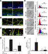
Figure 1.
Figure 1.
Occludin endocytosis mediates TNF-α–induced tight junction regulation in vivo. (A) Wild-type (WT) mice received TNF-α by i.p. injection and jejunal segments were harvested and stained for occludin (green), F-actin (red), and nuclei (blue) at designated times. Note the onset of occludin endocytosis at 90 min after TNF-α administration. Bar, 10 μm. (B) When viewed en face, it is clear that, focally, TNF-α induced complete depletion of tight junction-associated occludin. Bar, 10 μm. (C) Intestinal epithelial-specific occludin overexpression (OCLN Tg) prevents depletion of tight junction-associated occludin at 120 min after TNF-α administration. Compare to areas of perijunctional F-actin (red) that lack occludin (green) in WT mice (arrows). Bar, 10 μm. (D) In vivo perfusion assays show that TNF-α–induced paracellular leakage of BSA from the bloodstream to the intestinal lumen is markedly reduced by transgenic occludin overexpression. (E) Electron micrographs of jejunum, morphometric analyses, and Gaussian fits show a marked increase in numbers of 80 nm cytoplasmic vesicles 90 min after TNF-α injection. The decrease in 80 nm cytoplasmic vesicle numbers at 120 min coincides with increase numbers of larger vesicles, suggesting endosomal maturation. Bar, 500 nm. (F) In vivo perfusion data show that paracellular BSA leakage is not increased by TNF-α in caveolin-1 gene knockout (Cav1−/−) mice. Originally published in Marchiando et al.
Figure 2.
Figure 2.
Occludin expression confers colitis sensitivity by enhancing caspase-3 expression. (A) Occludin gene knockout (KO) attenuates DSS-induced weight loss. Sensitivity of Ocln knockout mice is restored to that of wild-type mice (WT) by transgenic intestinal epithelial occludin expression (KO/Tg). (B) Apoptotic ISOL-positive cells (red) numbers are increased after DSS challenge in WT, but not intestinal epithelial–specific Ocln knockout (KOIEC) mice. Nuclei are shown in blue for reference. Bar = 20 μm. (C) 5-Fluoruracil induces epithelial apoptosis as detected by cleaved caspase-3 staining (red) in WT, but not occludin KO, mice. Bar = 20 μm. (D) TNF-α induces caspase-3 cleavage and apoptosis in WT, but not Ocln KO, mice. Bar = 50 μm. (E) Quantitative qRT-PCR demonstrates reduced Casp3 mRNA in jejunal epithelial cells from occludin KO, relative to WT, mice. (F) Caspase-3 protein expression is reduced in jejunal epithelial cells isolated from Ocln KO mice. Caspase-8, caspase-9, and E-cadherin expression are not affected by occludin deletion. (G) Occludin promotes apoptosis by enhancing caspase-3 transcription, suggesting that occludin downregulation in inflammatory conditions may confer apoptotic resistance and promote mucosal homeostasis. Originally published in Kuo et al.
Figure 3.
Figure 3.
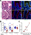
Occludin downregulation in inflammatory bowel disease is accompanied by reduced caspase-3 expression. (A) Hematoxylin-eosin (H&E) and immunofluorescent staining of occludin (green) and caspase-3 (green) in ileal biopsies from healthy control subjects and Crohn’s disease patients. E-cadherin (red) and nuclei (blue) are shown for reference. Bar = 50 μm. (B) Quantitative morphometry of occludin, caspase-3, and E-cadherin staining intensity within the intestinal epithelium. (C) Caspase-3 expression correlates with occludin expression (r2 = 0.76). Originally published in Kuo et al.
Figure 4.
Figure 4.
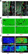
ZO-1 regulates cortical actomyosin and epithelial structure. (A) The smooth apical surface normally seen in wild-type (WT) epithelial monolayers is disrupted by apical distensions in jejunum from intestinal epithelial-specific ZO-1 gene knockout (KOIEC) mice. NHE3 (green), ZO-1 (red), E-cadherin (cyan), and nuclei (white). Bars = 100 μm (low magnification) and 10 μm (high magnification). (B) 3D reconstructions of F-actin–labeled epithelial surfaces highlight the apical distensions of KOIEC epithelium and show that blebbistatin, a myosin II inhibitor, normalizes apical contours. (C) The effect of apical distensions induced by in vitro ZO-1 knockdown (ZO-1KD) is emphasized in reconstructions by pseudocoloring each cell according to height (violet = 6 μm, red = 15 μm). Similar to the in vivo effect of blebbistatin, in vitro application of latrunculin A, which causes F-actin disruption, normalizes the height of ZO-1KD epithelial cells. Bars = 10 μm. Originally published in Odenwald et al.
Figure 5.
Figure 5.
ZO-1 directs epithelial growth and repair. (A) WT and ZO-1 knockdown (ZO-1KD) MDCK epithelial cells were stained for ZO-1 (red), E-cadherin (green), F-actin (purple), and nuclei (blue) at days 4, 8, and 11 after plating in collagen gels. Lumen formation (arrows) is delayed in ZO-1KD epithelial cells, which also fail to resolve the normally transient multilumen phase to form single-lumen cysts. Bars = 20 μm (low magnification) and 10 μm (high magnification). (B) Wildtype (WT, blue) and intestinal epithelial-specific ZO-1 gene knockout (KOIEC, red) mice were treated with DSS for 6 days. Weight loss is exaggerated and recovery is delayed in KOIEC mice. Bar = 50 μm. (C) One day after discontinuing DSS treatment (day 7), mice were injected with EdU (green) and sacrificed 2 hours later in order to track proliferating cells. Despite significant damage, the proliferative response of KOIEC mice is defective. (D) Gene set enrichment analysis (GSEA) of RNA-sequencing data from jejunal epithelia harvested 2 days after induction of cytokine storm (by anti-CD3 antibody injection). Activation of WNT–β-catenin, mitotic spindle, G2/M check point, and DNA repair pathways is deficient in KOIEC mice. (E) Colonoids from WT and KOIEC mice were treated with the GSK3β inhibitor CHIR99021 to potentiate WNT signaling and fixed 2 hours after EdU (green) addition. Numbers of EdU-labeled cells increased markedly in WT, but not KOIEC, colonoids. Bar = 20 μm (F) CHIR99021 induces extensive apoptosis in KOIEC, but not WT, colonoids. Bar = 20 μm. Panel A: Originally published in Odenwald et al. Panels B-F: Originally published in Kuo et al.
Figure 6.
Figure 6.
ZO-1 associates with spindle poles facilitating correct mitotic spindle orientation. (A) Live imaging of colonoids expressing EGFP-β-actin (magenta) and H2B-mCherry (cyan) shows that the mitotic spindle (arrows) is correctly oriented perpendicular to the basement membrane in wildtype (WT) but not ZO-1 gene knockout (KOIEC) cells. Drawings are presented to simplify interpretation. Bars = 10 μm. (B) Live imaging of mCherry-ZO-1 (green) expressing colonoids labeled with an intravital DNA stain (red) shows transient ZO-1 association with mitotic spindle poles (arrows). Bar = 5 μm. Drawings are presented to simplify interpretation. (C) Non-canonical functions, including regulation of WNT signaling and mitotic spindle orientation, make ZO-1 critical to mucosal repair. Reduced ZO-1 expression may contribute to ineffective mucosal healing in inflammatory bowel disease. Originally published in Kuo et al.

References

    1. Anderson JM & Van Itallie CM 2009. Physiology and function of the tight junction. Cold Spring Harb Perspect Biol. 1: a002584. - PMC - PubMed
    1. Van Itallie CM & Anderson JM 2006. Claudins and epithelial paracellular transport. Annu. Rev. Physiol 68: 403–429. - PubMed
    1. Turner JR 2009. Intestinal mucosal barrier function in health and disease. Nat Rev Immunol. 9: 799–809. - PubMed
    1. Shen L, Weber CR, Raleigh DR, Yu D & Turner JR 2011. Tight junction pore and leak pathways: A dynamic duo. Annu. Rev. Physiol 73: 283–309. - PMC - PubMed
    1. Marchiando AM, Graham WV & Turner JR 2010. Epithelial barriers in homeostasis and disease. Annu Rev Pathol. 5: 119–144. - PubMed

Publication types

MeSH terms