. 2022 May 17;12(1):8162.
doi: 10.1038/s41598-022-11382-7.

Helical Indexing in Real Space

Affiliations

Helical Indexing in Real Space

Chen Sun et al. Sci Rep. .

Abstract

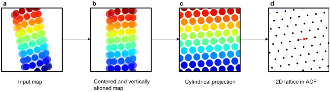
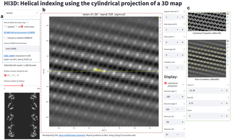
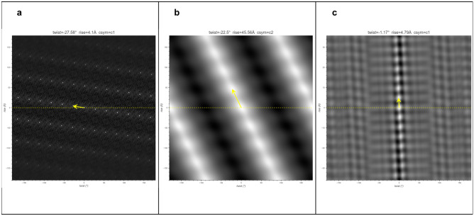
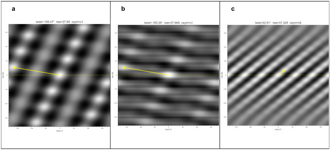
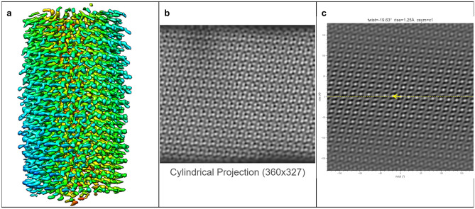
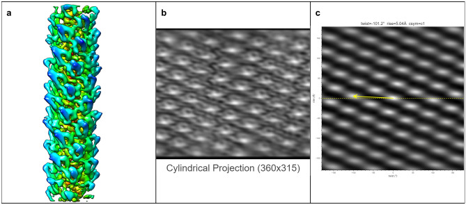
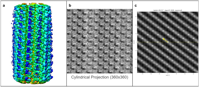
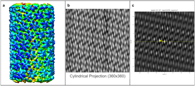
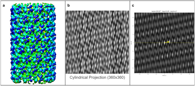
Biological structures with helical symmetries of distinct twist, rise, and axial symmetry are abundant and span a wide range of organisms and functions. Performing de novo helical indexing remains challenging because of the steep learning curve involved in Fourier space layer lines. The unknown amount of out-of-plane tilt and the existence of multiple conformations of the helices further complicate indexing. In this work, we introduce a real-space indexing method that leverages the prior knowledge of the tilt and in-plane angles of the helical filaments/tubes, robust ab initio 3D reconstruction capabilities in single particle cryo-EM to obtain asymmetric reconstructions, and automatic indexing of helical parameters directly from the asymmetric density maps. We validated this approach using data from multiple helical structures of distinct helical symmetries, diameters, flexibility, data qualities, and heterogeneous states. The fully automated tool we introduce for real space indexing, HI3D, uses the 2D lattice in the autocorrelation of the cylindrical projection of a 3D density map to identify the helical symmetry. HI3D can often successfully determine the helical parameters of a suboptimal 3D density map, including ab initio single particle asymmetric reconstructions and sub-tomogram averages, with intermediate evidence that can also help assess the map quality. Furthermore, this open-source HI3D is usable independently as a Web application that can be accessed free of installation. With these methods, de novo helical indexing will be significantly more accessible to researchers investigating structures of helical filaments/tubes using cryo-EM.


Conflict of interest statement

The authors declare no competing interests.

Figures

Figure 1
Figure 1
HI3D workflow. The input asymmetric density map (a) that is arbitrarily positioned and orientated is automatically centered and vertically aligned (b). The aligned 3D map is resampled in the cylindrical coordinate to generate the cylindrical projection (c) to mathematically convert a helical structure in the original 3D map into a “2D crystal” image. The auto-correlation function of the cylindrical projection would generate a 2D lattice (d) that visually resembles the diffraction spots of a crystal. The unit cell vector (red arrow in d) with the shortest distance to the equator would correspond to the helical twist (x-coordinate) and rise (y-coordinate).
Figure 2
Figure 2
HI3D Web app user interface. The user interface of HI3D consists of three major parts. (a) The left part shows the input panel and the X/Y/Z section of the input map. Users can input a map in three ways. The first one is to upload a map from the local directory. The second is to provide the URL, for example, of a cryoSPARC output map. The third is dedicated to the helical structures in EMDB by either entering an EMDB ID or randomly choosing a helical structure in EMDB. (b) The central panel shows the output twist, rise, and C-sym values in text and as a vector centered in the ACF image. The right panel (c) shows the cylindrical projection (top), ACF of the projection image (middle), and input fields (bottom) for the user to overwrite the rise and twist if the automated detection fails.
Figure 3
Figure 3
HI3D results for three helical structures in EMDB. (a) EMD-30129 (Helical stem of the cleaved double-headed nucleocapsids of Sendai virus, 2.9 Å); (b) EMD-1759 (Bacterial tubulin homologue TubZ, 35 Å, reconstructed from negative stain EM images); (c) EMD-23890 (Tau straight helical filament extracted from PrP-CAA patient brain tissue, 3.1 Å). The published rise and twist for these three datasets are (4.09 Å, -27.58), (42 Å, 21), and (4.79 Å, -1.07), respectively.
Figure 4
Figure 4
HI3D results for three subtomogram averages in EMDB. (a) EMD-12289 (in situ actomyosin, 10.2 Å); (b) EMD-12293 (in situ I-band thin filament including troponin complex, 19.8 Å); (c) EMD-8601 (in situ type VI secretion system, 24 Å).
Figure 5
Figure 5
The surface view of the asymmetric reconstruction (a), the cylindrical projection (b), and the 2D lattice (c) with the HI3D outputted helical rise and twist and C-sym of the TMV dataset (EMPIAR-1022). The surface view was generated with UCSF Chimera.
Figure 6
Figure 6
The surface view of the asymmetric reconstruction (a), the cylindrical projection (b), and the 2D lattice (c) with the HI3D outputted helical rise and twist and C-sym of the MAVS CARD dataset (EMPIAR-10031). The surface view was generated with UCSF Chimera.
Figure 7
Figure 7
The surface view of the asymmetric reconstruction (a), the cylindrical projection (b), and the 2D lattice (c) with the HI3D outputted helical rise and twist and C-sym of the VipA/VipB dataset (EMPIAR-10029). The surface view was generated with UCSF Chimera.
Figure 8
Figure 8
The surface view of the asymmetric reconstruction (a), the cylindrical projection (b), and the 2D lattice (c) with the HI3D outputted helical rise and twist and C-sym of the major class of the HIV capsid protein dataset (provided by Dr. Peijun Zhang). The surface view was generated with UCSF Chimera.
Figure 9
Figure 9
The surface view of the asymmetric reconstruction (a), the cylindrical projection (b), and the 2D lattice (c) with the HI3D outputted helical rise and twist and C-sym of the minor class of the HIV capsid protein dataset (provided by Dr. Peijun Zhang). The surface view was generated with UCSF Chimera.

Similar articles

Cited by

References

    1. Lynch EM, Kollman JM, Webb BA. Filament formation by metabolic enzymes-a new twist on regulation. Curr. Opin. Cell Biol. 2020;66:28–33. doi: 10.1016/j.ceb.2020.04.006. - DOI - PMC - PubMed
    1. Berriman J, et al. Tau filaments from human brain and from in vitro assembly of recombinant protein show cross-beta structure. Proc. Natl. Acad. Sci. USA. 2003;100:9034–9038. doi: 10.1073/pnas.1530287100. - DOI - PMC - PubMed
    1. Kidd, M. Paired helical filaments in electron microscopy of alzheimer’s disease (1963). - PubMed
    1. Terry, R. D. The fine structure of neurofibrillary tangles in Alzheimer’s disease (1963). - PubMed
    1. De Rosier, D. J. & Klug, A. Reconstruction of three dimensional structures from electron micrographs (1968). - PubMed

Publication types

MeSH terms