

The effect of melatonin on the mouse ameloblast-lineage cell line ALCs
- PMID: 35581244
- PMCID: PMC9114102
- DOI: 10.1038/s41598-022-11912-3
The effect of melatonin on the mouse ameloblast-lineage cell line ALCs
Abstract
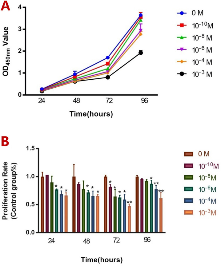
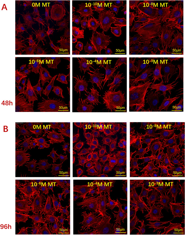
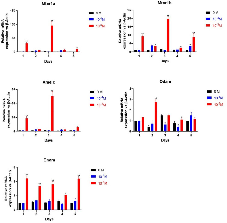
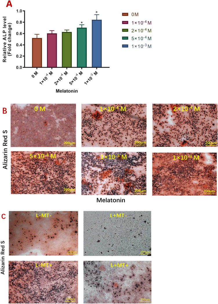
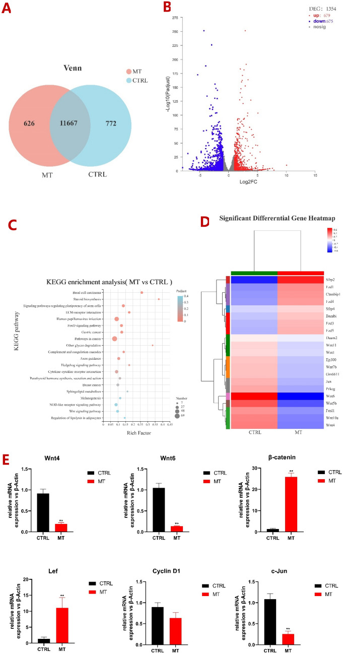
Melatonin plays a critical role in promoting the proliferation of osteoblasts and the growth and development of dental papilla cells. However, the effect and mechanism of melatonin on the growth and development of ALCs still need to be explored. CCK8 assay was used for the evaluation of cell numbers. qRT-PCR was used to identify the differentially expressed genes in ALCs after melatonin treatment. The number and morphology of ALCs were investigated by confocal microscopy. Alkaline phosphatase assay and Alizarin red S staining were used for measuring mineralization. Then, we focused on observing the crucial factors of the signaling pathway by RNA-seq and qRT-PCR. Melatonin limited the cell number of ALCs in a dose-dependent manner and promoted the production of actin fibers. A high concentration of melatonin significantly promoted the mRNA levels of enamel matrix proteins and the formation of mineralized nodules. RNA-seq data showed that Wnt signaling pathway may be involved in the differentiation of ALCs under the influence of melatonin. This study suggests that melatonin plays a regulatory role in the cell number, differentiation, and mineralization of the ALCs, and then shows the relationship between the Wnt signaling pathway with the ALCs under melatonin.
© 2022. The Author(s).
Conflict of interest statement
The authors declare no competing interests.
Figures
Similar articles
-
Melatonin-Medicated Neural JNK3 Up-Regulation Promotes Ameloblastic Mineralization.Front Cell Dev Biol. 2021 Dec 24;9:749642. doi: 10.3389/fcell.2021.749642. eCollection 2021. Front Cell Dev Biol. 2021. PMID: 35004671 Free PMC article.
-
Role of protein delta homolog 1 in the proliferation and differentiation of ameloblasts.Mol Med Rep. 2018 Mar;17(3):3537-3544. doi: 10.3892/mmr.2017.8290. Epub 2017 Dec 18. Mol Med Rep. 2018. PMID: 29257328 Free PMC article.
-
Melatonin rescues glucocorticoid-induced inhibition of osteoblast differentiation in MC3T3-E1 cells via the PI3K/AKT and BMP/Smad signalling pathways.Life Sci. 2020 Sep 15;257:118044. doi: 10.1016/j.lfs.2020.118044. Epub 2020 Jul 2. Life Sci. 2020. PMID: 32622944
-
Small molecules direct the generation of ameloblast-like cells from human embryonic stem cells.Stem Cell Res Ther. 2025 Apr 12;16(1):173. doi: 10.1186/s13287-025-04294-6. Stem Cell Res Ther. 2025. PMID: 40221796 Free PMC article.
-
Melatonin promotes osteoblastic differentiation and regulates PDGF/AKT signaling pathway.Cell Biol Int. 2020 Feb;44(2):402-411. doi: 10.1002/cbin.11240. Epub 2019 Oct 2. Cell Biol Int. 2020. PMID: 31535749
Cited by
-
Neural Regulations in Tooth Development and Tooth-Periodontium Complex Homeostasis: A Literature Review.Int J Mol Sci. 2022 Nov 16;23(22):14150. doi: 10.3390/ijms232214150. Int J Mol Sci. 2022. PMID: 36430624 Free PMC article. Review.
-
Melatonin pretreatment on exosomes: Heterogeneity, therapeutic effects, and usage.Front Immunol. 2022 Sep 16;13:933736. doi: 10.3389/fimmu.2022.933736. eCollection 2022. Front Immunol. 2022. PMID: 36189281 Free PMC article. Review.
-
Melatonin ameliorates inflammation-induced developmental defects of enamel by upregulating regulator of G protein signaling 2.J Dent Sci. 2024 Oct;19(4):2355-2366. doi: 10.1016/j.jds.2024.01.019. Epub 2024 Feb 6. J Dent Sci. 2024. PMID: 39347090 Free PMC article.
References
Publication types
MeSH terms
Substances
LinkOut - more resources
Full Text Sources
