. 2022 May 17;12(1):8165.
doi: 10.1038/s41598-022-12057-z.

Cell-controlled dynamic surfaces for skeletal stem cell growth and differentiation

Affiliations

Cell-controlled dynamic surfaces for skeletal stem cell growth and differentiation

Hilary J Anderson et al. Sci Rep. .

Abstract

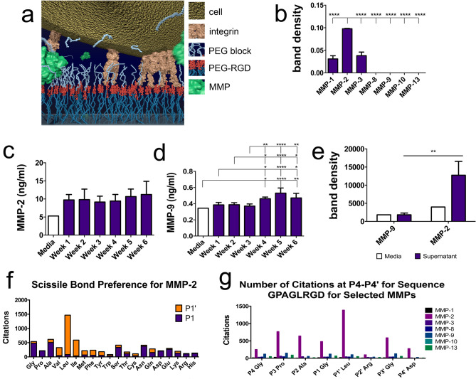
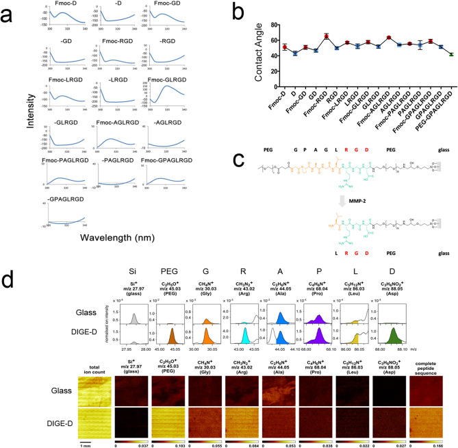
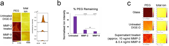
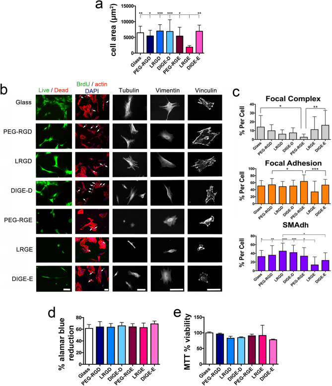
Skeletal stem cells (SSCs, or mesenchymal stromal cells typically referred to as mesenchymal stem cells from the bone marrow) are a dynamic progenitor population that can enter quiescence, self-renew or differentiate depending on regenerative demand and cues from their niche environment. However, ex vivo, in culture, they are grown typically on hard polystyrene surfaces, and this leads to rapid loss of the SSC phenotype. While materials are being developed that can control SSC growth and differentiation, very few examples of dynamic interfaces that reflect the plastic nature of the stem cells have, to date, been developed. Achieving such interfaces is challenging because of competing needs: growing SSCs require lower cell adhesion and intracellular tension while differentiation to, for example, bone-forming osteoblasts requires increased adhesion and intracellular tension. We previously reported a dynamic interface where the cell adhesion tripeptide arginine-glycine-aspartic acid (RGD) was presented to the cells upon activation by user-added elastase that cleaved a bulky blocking group hiding RGD from the cells. This allowed for a growth phase while the blocking group was in place and the cells could only form smaller adhesions, followed by an osteoblast differentiation phase that was induced after elastase was added which triggered exposure of RGD and subsequent cell adhesion and contraction. Here, we aimed to develop an autonomous system where the surface is activated according to the need of the cell by using matrix metalloprotease (MMP) cleavable peptide sequences to remove the blocking group with the hypothesis that the SSCs would produce higher levels of MMP as the cells reached confluence. The current studies demonstrate that SSCs produce active MMP-2 that can cleave functional groups on a surface. We also demonstrate that SSCs can grow on the uncleaved surface and, with time, produce osteogenic marker proteins on the MMP-responsive surface. These studies demonstrate the concept for cell-controlled surfaces that can modulate adhesion and phenotype with significant implications for stem cell phenotype modulation.


Conflict of interest statement

The authors declare no competing interests.

Figures

Figure 1
Figure 1
Selecting MMPs and cleavage sequence. (a) Schematic of the SSC cleavable surface concept with MMP-driven removal of PEG revealing RGD to the cells. (b) After 3 weeks of culture, MMP protein array was performed by densitometry measurements of the array membranes demonstrating high levels of MMP-2 expression (n = 3). (c,d) ELISA showed constant expression of MMP-2 and increasing expression of MMP-9 with time (n = 3). (e) Zymography was used to show that only MMP-2 was expressed by the SSCs in active form after 3 weeks of culture (n = 3). (f) MERPOS data gave consensus of an 8-amino acid sequence with a strong preference for L at P1′; allowing us to design a GPAG↓LRGD sequence for MMP-2 cleavage that would leave RGD on the coverslip. (g) GPAG↓LRGD sequence is highly cited as being MMP responsive For (b–e), results are mean ± SD, statistics by ANOVA and Tukey test where *p < 0.05, **p < 0.01, ***p < 0.001 and ****p < 0.0001. If stars are not shown on the graphs in (b–e), it denotes no significant difference was observed between the treatment and relevant control.
Figure 2
Figure 2
Characterisation of solid-phase peptide synthesis (SPPS) dynamic surface. (a) Surface fluorescence spectroscopy following addition/removal of the Fmoc group (Fmoc peak seen at ~ 315 nm) during synthesis. (b) Contact angle measurement following addition (red)/removal (blue) of the Fmoc group during synthesis and the final PEG-capped peptide sequence (green) (graph shows mean ± SD, 50 images per dataset with 15 datasets taken across 3 substrates (n = 3)). (c) Scheme showing complete RGD surface with MMP cleavable group and PEG blocker before and after MMP cleavage. (d) Tof–SIMS data showing that expected amino acids (G, P, A, L, R and D) and PEG were present post-synthesis. Together, this data indicates complete PEG-GPAGLRGD synthesis.
Figure 3
Figure 3
MMPs can cleave PEG from the peptide and reveal RGD. (a,b) Tof–SIMS data showing removal of the PEG group with the addition of MMPs-2 and 9 in more detail. (c) Treatment of the DIGE-D surface with SSC supernatant containing 10 ng/mL of MMP-2 (active) and 0.4 ng/mL of MMP-9 (latent) resulted in some cleavage of PEG.
Figure 4
Figure 4
SSC biocompatibility. (a) Cell area measurements after 24 h of culture on the samples showing that RGD containing surfaces and DIGE-E supported similar cell spreading compared to glass while LRGE, where the RGE was exposed, reduced SSC spreading (n = 15). (b) Viability, proliferation and cytoskeletal analysis showed high cell viability on all surfaces. Growth and cytoskeletal organisation were reduced on the LRGE surface and PEG-RGE surface where the RGE group was exposed. (c) Focal adhesions were categorised as FC (< 2 µm), FA (2–5 µm) and SMAdh (> 5 µm) and expressed as % total adhesions (n = 15). The main difference to indicate cells responding to the PEG cleavage was seen in the SMAdhs, where LRGD and DIGE-D gave the largest differences to the RGE containing surfaces. (d) Alamar blue and MTT metabolic assays showing no differences (n = 3). Graphs show mean ± SD, statistics by ANOVA and Tukey test where *p < 0.05, **p < 0.01, ***p < 0.001 and ****p < 0.0001. If stars are not shown on the graphs, it denotes no significant difference was observed between the treatment and relevant control. Please note that SSCs purchased from Promocell GMBH rather than stro-1+ve cells were used to create this figure.
Figure 5
Figure 5
SSC phenotype on DIGE-D surface. (a) In cell western (ICW) data for Stro-1 showing no difference between SSCs cultured on glass control and DIGE-D and DIG-E surfaces showing that cells express similar levels of this SSC marker on the SPPS test and control surfaces when the RGD/RGE is hidden by the PEG blocking group. (b,c) ICW data for alkaline phosphatase (ALP), osteopontin (OPN) and osteocalcin (OCN) after 4 weeks (b) and 6 weeks (c) of culture. No differences were noted between samples at 4 weeks, but at 6 weeks, higher levels of OCN expression were noted in SSCs on the DIGE-D surface. (d) QPCR for OPN transcripts after 4 and 6 weeks of MSC culture on PEG, LRGD and DIGE-D surfaces. At 4 weeks no change was seen. However, at 6 weeks, the DIGE-D MMP-responsive surface supported significantly greater OPN expression. Graphs show mean ± SD, n = 3, statistics by ANOVA and Tukey test where *p < 0.05, **p < 0.01, ***p < 0.001 and ****p < 0.0001. If stars are not shown on the graphs, it denotes no significant difference was observed between the treatment and relevant control.

Similar articles

Cited by

References

    1. Mirmalek-Sani SH, et al. Characterization and multipotentiality of human fetal femur-derived cells—Implications for skeletal tissue regeneration. Stem Cells. 2006;24:1042–1053. doi: 10.1634/stemcells.2005-0368. - DOI - PubMed
    1. Bianco P, Riminucci M, Gronthos S, Robey PG. Bone marrow stromal stem cells: Nature, biology, and potential applications. Stem Cells. 2001;19:180–192. doi: 10.1634/stemcells.19-3-180. - DOI - PubMed
    1. Tome M, Lindsay SL, Riddell JS, Barnett SC. Identification of nonepithelial multipotent cells in the embryonic olfactory mucosa. Stem Cells. 2009;27:2196–2208. doi: 10.1002/stem.130. - DOI - PubMed
    1. Watt FM, Hogan BL. Out of Eden: Stem cells and their niches. Science. 2000;287:1427–1430. doi: 10.1126/science.287.5457.1427. - DOI - PubMed
    1. Yang C, Tibbitt MW, Basta L, Anseth KS. Mechanical memory and dosing influence stem cell fate. Nat. Mater. 2014;13:645–652. doi: 10.1038/nmat3889. - DOI - PMC - PubMed

Publication types