

CT-based radiomics signature for differentiation between cardiac tumors and a thrombi: a retrospective, multicenter study
- PMID: 35581366
- PMCID: PMC9114026
- DOI: 10.1038/s41598-022-12229-x
CT-based radiomics signature for differentiation between cardiac tumors and a thrombi: a retrospective, multicenter study
Abstract
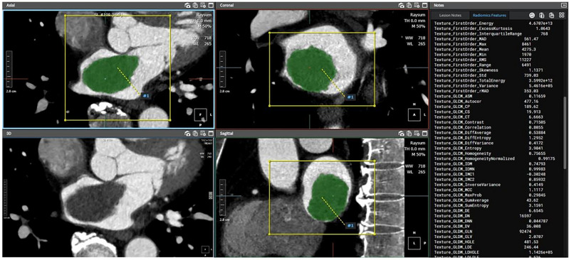
The study aimed to develop and validate whether the computed tomography (CT) radiomics analysis is effective in differentiating cardiac tumors and thrombi. For this retrospective study, a radiomics model was developed on the basis of a training dataset of 192 patients (61.9 ± 13.3 years, 90 men) with cardiac masses detected in cardiac CT from January 2010 to September 2019. We constructed three models for discriminating between a cardiac tumor and a thrombus: a radiomics model, a clinical model, which included clinical and conventional CT variables, and a model that combined clinical and radiomics models. In the training dataset, the radiomics model and the combined model yielded significantly higher differentiation performance between cardiac tumors and cardiac thrombi than the clinical model (AUC 0.973 vs 0.870, p < 0.001 and AUC 0.983 vs 0.870, p < 0.001, respectively). In the external validation dataset with 63 patients (59.8 ± 13.2 years, 26 men), the combined model yielded a larger AUC compared to the clinical model (AUC 0.911 vs 0.802, p = 0.037). CT radiomics analysis is effective in differentiating cardiac tumors and thrombi. In conclusion, the combination of clinical, conventional CT, and radiomics features demonstrated an additional benefit in differentiating between cardiac tumor and thrombi compared to clinical data and conventional CT features alone.
© 2022. The Author(s).
Conflict of interest statement
The authors declare no competing interests.
Figures


Similar articles
-
Distinguishing cardiac myxomas from cardiac thrombi by a radiomics signature based on cardiovascular contrast-enhanced computed tomography images.BMC Cardiovasc Disord. 2021 Mar 25;21(1):152. doi: 10.1186/s12872-021-01961-3. BMC Cardiovasc Disord. 2021. PMID: 33765929 Free PMC article.
-
A CT-based radiomics nomogram for differentiation of renal angiomyolipoma without visible fat from homogeneous clear cell renal cell carcinoma.Eur Radiol. 2020 Feb;30(2):1274-1284. doi: 10.1007/s00330-019-06427-x. Epub 2019 Sep 10. Eur Radiol. 2020. PMID: 31506816
-
Radiomics analysis of dual-energy CT-derived iodine maps for diagnosing metastatic cervical lymph nodes in patients with papillary thyroid cancer.Eur Radiol. 2020 Nov;30(11):6251-6262. doi: 10.1007/s00330-020-06866-x. Epub 2020 Jun 4. Eur Radiol. 2020. PMID: 32500193
-
Radiomics analysis of multicenter CT images for discriminating mucinous adenocarcinoma from nomucinous adenocarcinoma in rectal cancer and comparison with conventional CT values.J Xray Sci Technol. 2020;28(2):285-297. doi: 10.3233/XST-190614. J Xray Sci Technol. 2020. PMID: 32116286
-
Clot-based radiomics model for cardioembolic stroke prediction with CT imaging before recanalization: a multicenter study.Eur Radiol. 2023 Feb;33(2):970-980. doi: 10.1007/s00330-022-09116-4. Epub 2022 Sep 6. Eur Radiol. 2023. PMID: 36066731
Cited by
-
A Novel Data Augmentation Method for Radiomics Analysis Using Image Perturbations.J Imaging Inform Med. 2024 Oct;37(5):2401-2414. doi: 10.1007/s10278-024-01013-0. Epub 2024 May 6. J Imaging Inform Med. 2024. PMID: 38710969 Free PMC article.
-
Cardiac Masses and Pseudomasses: An Overview about Diagnostic Imaging and Clinical Background.Medicina (Kaunas). 2023 Dec 29;60(1):70. doi: 10.3390/medicina60010070. Medicina (Kaunas). 2023. PMID: 38256331 Free PMC article. Review.
-
Clinical characteristics of primary atrial tumor and their diagnostic value: A retrospective study of 10 years.Front Surg. 2023 Feb 14;10:1097287. doi: 10.3389/fsurg.2023.1097287. eCollection 2023. Front Surg. 2023. PMID: 36865623 Free PMC article.
-
The diagnostic accuracy of contrast echocardiography in patients with suspected cardiac masses: A preliminary multicenter, cross-sectional study.Front Cardiovasc Med. 2022 Sep 16;9:1011560. doi: 10.3389/fcvm.2022.1011560. eCollection 2022. Front Cardiovasc Med. 2022. PMID: 36187014 Free PMC article.
-
The usefulness of contrast echocardiography in the evaluation of cardiac masses: a multicenter study.BMC Cardiovasc Disord. 2024 Jan 13;24(1):43. doi: 10.1186/s12872-024-03708-2. BMC Cardiovasc Disord. 2024. PMID: 38218809 Free PMC article.
References
Publication types
MeSH terms
LinkOut - more resources
Full Text Sources
Medical
