

Deep Learning Achieves Neuroradiologist-Level Performance in Detecting Hydrocephalus Requiring Treatment
- PMID: 35581409
- PMCID: PMC9712867
- DOI: 10.1007/s10278-022-00654-3
Deep Learning Achieves Neuroradiologist-Level Performance in Detecting Hydrocephalus Requiring Treatment
Abstract
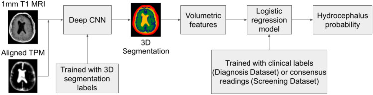
In large clinical centers a small subset of patients present with hydrocephalus that requires surgical treatment. We aimed to develop a screening tool to detect such cases from the head MRI with performance comparable to neuroradiologists. We leveraged 496 clinical MRI exams collected retrospectively at a single clinical site from patients referred for any reason. This diagnostic dataset was enriched to have 259 hydrocephalus cases. A 3D convolutional neural network was trained on 16 manually segmented exams (ten hydrocephalus) and subsequently used to automatically segment the remaining 480 exams and extract volumetric anatomical features. A linear classifier of these features was trained on 240 exams to detect cases of hydrocephalus that required treatment with surgical intervention. Performance was compared to four neuroradiologists on the remaining 240 exams. Performance was also evaluated on a separate screening dataset of 451 exams collected from a routine clinical population to predict the consensus reading from four neuroradiologists using images alone. The pipeline was also tested on an external dataset of 31 exams from a 2nd clinical site. The most discriminant features were the Magnetic Resonance Hydrocephalic Index (MRHI), ventricle volume, and the ratio between ventricle and brain volume. At matching sensitivity, the specificity of the machine and the neuroradiologists did not show significant differences for detection of hydrocephalus on either dataset (proportions test, p > 0.05). ROC performance compared favorably with the state-of-the-art (AUC 0.90-0.96), and replicated in the external validation. Hydrocephalus cases requiring treatment can be detected automatically from MRI in a heterogeneous patient population based on quantitative characterization of brain anatomy with performance comparable to that of neuroradiologists.
Keywords: Brain MRIs; Convolutional neural networks; Deep learning; Hydrocephalus.
© 2022. The Author(s) under exclusive licence to Society for Imaging Informatics in Medicine.
Conflict of interest statement
On behalf of all authors, the corresponding author states that there is no relevant conflict of interest or industry support for the project. R. J. Y. has received research funding from Agios, and performed consulting for Agios, Puma, NordicNeuroLab, and ICON plc, all unrelated to the current work.
Figures


References
-
- Ishii K, Kanda T, Harada A, Miyamoto N, Kawaguchi T, Shimada K, et al. Clinical impact of the callosal angle in the diagnosis of idiopathic normal pressure hydrocephalus. Eur Radiol. 2008 May 24 [cited 2021 Oct 28];18(11):2678. Available from: 10.1007/s00330-008-1044-4 - PubMed
-
- Ambarki K, Israelsson H, Wåhlin A, Birgander R, Eklund A, Malm J. Brain ventricular size in healthy elderly: comparison between Evans index and volume measurement. Neurosurgery. 2010 Jul;67(1):94–9; discussion 99. - PubMed
-
- Yamada S, Ishikawa M, Yamamoto K. Optimal Diagnostic Indices for Idiopathic Normal Pressure Hydrocephalus Based on the 3D Quantitative Volumetric Analysis for the Cerebral Ventricle and Subarachnoid Space. American Journal of Neuroradiology. 2015 Dec 1 [cited 2020 Nov 27];36(12):2262–9. Available from: http://www.ajnr.org/content/36/12/2262 - PMC - PubMed
Publication types
MeSH terms
Grants and funding
LinkOut - more resources
Full Text Sources
Medical
