. 2022 May 18;20(1):116.
doi: 10.1186/s12915-022-01303-5.

Identification of LINE retrotransposons and long non-coding RNAs expressed in the octopus brain

Affiliations

Identification of LINE retrotransposons and long non-coding RNAs expressed in the octopus brain

Giuseppe Petrosino et al. BMC Biol. .

Abstract

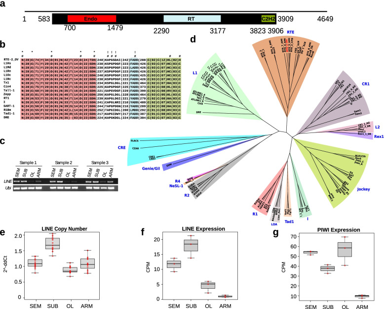
Background: Transposable elements (TEs) widely contribute to the evolution of genomes allowing genomic innovations, generating germinal and somatic heterogeneity, and giving birth to long non-coding RNAs (lncRNAs). These features have been associated to the evolution, functioning, and complexity of the nervous system at such a level that somatic retrotransposition of long interspersed element (LINE) L1 has been proposed to be associated to human cognition. Among invertebrates, octopuses are fascinating animals whose nervous system reaches a high level of complexity achieving sophisticated cognitive abilities. The sequencing of the genome of the Octopus bimaculoides revealed a striking expansion of TEs which were proposed to have contributed to the evolution of its complex nervous system. We recently found a similar expansion also in the genome of Octopus vulgaris. However, a specific search for the existence and the transcription of full-length transpositionally competent TEs has not been performed in this genus.

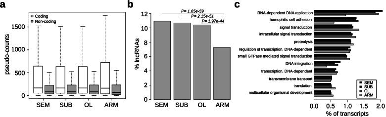
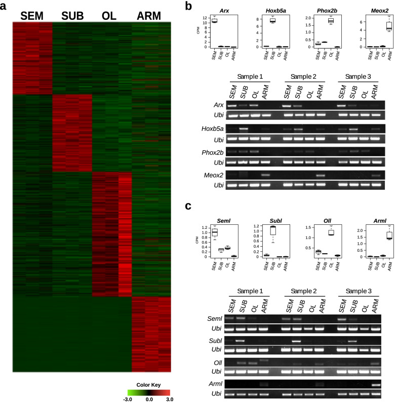
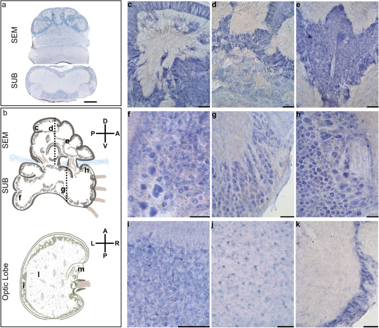
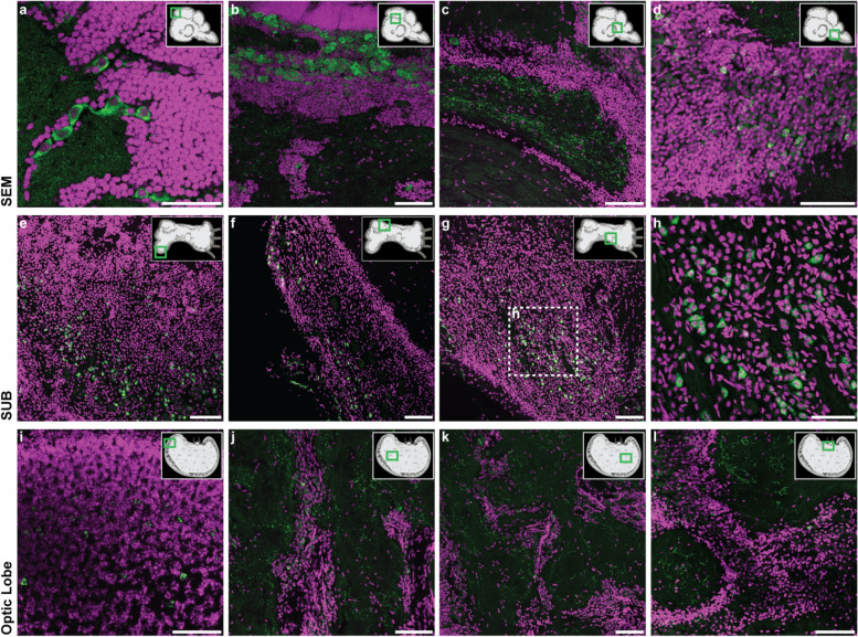
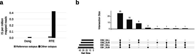
Results: Here, we report the identification of LINE elements competent for retrotransposition in Octopus vulgaris and Octopus bimaculoides and show evidence suggesting that they might be transcribed and determine germline and somatic polymorphisms especially in the brain. Transcription and translation measured for one of these elements resulted in specific signals in neurons belonging to areas associated with behavioral plasticity. We also report the transcription of thousands of lncRNAs and the pervasive inclusion of TE fragments in the transcriptomes of both Octopus species, further testifying the crucial activity of TEs in the evolution of the octopus genomes.

Conclusions: The neural transcriptome of the octopus shows the transcription of thousands of putative lncRNAs and of a full-length LINE element belonging to the RTE class. We speculate that a convergent evolutionary process involving retrotransposons activity in the brain has been important for the evolution of sophisticated cognitive abilities in this genus.

Keywords: Mollusks; Nervous system; Transcriptome; Transposable elements.


Conflict of interest statement

The authors declare that they have no competing interests.

Figures

Fig. 1
Fig. 1
Features of the Octopus vulgaris brain and arm transcriptome. We sequenced the supra-esophageal (SEM) and sub-esophageal (SUB) masses and optic lobe (OL) as representatives of the brain and the medial segment of an arm (ARM), including the arm nerve cord, as the representative of the peripheral system. a Expression levels for coding and non-coding transcripts. Non-coding transcripts are on average expressed at lower levels than coding. b Percentage of the expressed non-coding transcripts. Brain sample results enriched for non-coding. c Percentages of transcripts expressed and their relative distribution among the most represented GO biological processes. A higher percentage of transcripts belonging to classes related to transposable elements and cell adhesion are expressed in the brain. Transcripts likely to be involved in signal transduction and translation constitute a larger quota in the arm
Fig. 2
Fig. 2
Transcripts with an expression peak and transcriptome validations. a Heatmap showing the expression levels for transcripts classified as having a peak of expression. b Boxplots showing the RNAseq expression levels of coding transcripts selected for validation and their relative RT-PCR results from 3 different individuals. c Boxplots showing the RNAseq expression levels of non-coding transcripts selected for validation and their relative RT-PCR results from 3 different individuals. The octopus ubiquitin transcript (Ubi) has been used as a positive control in all the experiments
Fig. 3
Fig. 3
A full-length potentially active LINE transcribed in Octopus vulgaris. a Domain composition of the discovered LINE (comp36575_c1_seq3). The black line represents the transcript, the black box represents the location of the ORF, and the colored boxes represent the protein domains (Endo, endonuclease; RT, reverse transcriptase; C2H2, zinc finger). The numbers are relative to the nucleotide positions in the transcript. b Schematic alignment highlighting the conserved catalytic amino acids in the group of LINEs adapted from [38] plus the octopus element. The color code of the domains is the same as in a; amino acids critical for EN (*), RT (!), and retrotrasposition (#). c Electrophoresis from RT-PCR of the LINE showing the expression from three different animals. d Phylogenetic tree based on 100 LINEs from [39] (see Additional file 1: Table S4) plus the octopus element in red. e LINE copy number variation analysis using quantitative real-time PCR with Taqman probes. f Expression levels of the LINE based on RNAseq data. g Expression levels of Piwi-like protein 1 (comp33731_c0_seq1) from RNAseq data
Fig. 4
Fig. 4
Localization of RTE-2_OV mRNA by in situ hybridization. a Bright-field micrographs of the coronal sections of the supraesophageal (SEM) and subesophageal (SUB) masses hybridized to digoxigenin-labeled LINE antisense (refer to the plane indicated by the dashed lines in b). The five gyri of the vertical lobe (dorsal in SEM) appear positively marked by the RTE-2_OV mRNA. Several cells in the cortical layers of the SUB appear also stained after in situ hybridization. Neuropils of SEM and SUB do not show a signal. b Schematic outline of the parts of the octopus brain for SEM and SUB (sagittal plane) and the optic lobe (OL; horizontal plane). Axes illustrating dorso-ventral and antero-posterior (SEM and SUB) and antero-posterior and left-right (OL) orientations with respect to the octopus body plan. Black letters indicate approximate levels of the sections provided in the other panels of the figure. c Detail of a gyrus of the vertical lobe (SEM) with densely packed amacrine cells showing a positive signal. d A similar signal in the gyri of the vertical lobe and some scattered positive cells in the sub-vertical lobe. e Section at the level of the sub-frontal lobe with densely packed amacrine small cells showing a strong positive signal. In the SUB, we observed f positive cells (20–25 μm in diameter) in the pallovisceral lobe and some larger neurons (40–50 μm) belonging to typical motor-center cellular types. g Cells (20–25 μm) belonging to the ventral side of the anterior pedal lobe and at the level of the dorsal brachial lobe (h) where some larger cells (up to 50 μm) are also marked after ISH. The small cells pertaining to these areas do not show positivity. Details of horizontal sections of the O. vulgaris optic lobe (i, j, k; in areas indicated in b): i Outer layer rich in intensely positive cells (small amacrine cells, < 5 μm), j inner medulla with scattered LINE mRNA-expressing cell bodies (up to 10 μm), and k cell bodies of the peduncle complex at the level of the median and posterior lobules of the olfactory lobe (cells of about 10 μm). Scale bars, 100 μm and 500 μm in a. Schematic drawings in b not to scale
Fig. 5
Fig. 5
RTE-2_OV immunostaining in different areas of the brain. Coronal sections of the supra-esophageal (SEM; ad) and sub-esophageal (SUB; eh) masses and horizontal sections for the optic lobe (OL; il) following fluorescent-IHC (RTE-2_OV signal in green, DAPI used as a nuclear stain in magenta) highlight a differential pattern of positive cells and fibers in O. vulgaris brain. A schematic drawing of the brain parts is provided with areas of interest indicated in the green square. a Large positive cells are found in the vertical lobe (VL). These appear organized in trunks and clearly distinguishable from the population of numerous amacrine cells constituting the VL (DAPI stained layer). b Large cells in the sub-vertical lobe (cellular wall) and a part of the bundle of fibers are present at the beginning of the sub-frontal lobe (c). d Scattered positive cells are also identified in the posterior buccal lobe. Several positive cells are identified in the SUB in the cellular walls of the vasomotor lobe (e) and in discrete areas of the pedal lobe (f). A similar pattern of positive cells is recognized at the level of the anterior part of the pedal lobe (g). A detail in the higher magnification (h; square in g) of the cellular layer of the lobe serves to highlight the population of positive cells. In the OL, several amacrine cells are found positive in the external granular layer (i). The OL-medulla is populated by few immune-reactive neurons found in the cellular islands (j, k), and positive fibers dispersed in the surrounding neuropil (j, k). Positive cells and fibers are also identified in the peduncle lobe. The internal layer of cells of the neural wall of the olfactory lobe and the spine (l) are shown. Scale bars, 100 μm, with the exception of a and h (50 μm)
Fig. 6
Fig. 6
Identification of a putatively active LINE transcribed in O. bimaculoides. a Normalized number of non-reference insertions in two different O. bimaculoides individuals (octopus used for the reference genome and a different octopus individual) for the two LINEs identified in this species. b Upset plot showing the non-reference insertions identified in the 30X WGS from two different samples from 2 different individuals and how they are shared between the four samples. OB1_SUB: sample from the subesophageal mass of the octopus 1. OB1_GILL, sample from the gill of octopus 1; OB2_SUB, sample from the subesophageal mass of the octopus 2; OB2_GILL, sample from the gill of octopus 2

Similar articles

Cited by

References

    1. Kazazian HH. Mobile elements: drivers of genome evolution. Science. 2004;303:1626–1632. doi: 10.1126/science.1089670. - DOI - PubMed
    1. Kelley DR, Rinn JL. Transposable elements reveal a stem cell specific class of long noncoding RNAs. Genome Biol. 2012;13:R107. doi: 10.1186/gb-2012-13-11-r107. - DOI - PMC - PubMed
    1. Kapusta A, Kronenberg Z, Lynch VJ, Zhuo X, Ramsay L, Bourque G, et al. Transposable elements are major contributors to the origin, diversification, and regulation of vertebrate long noncoding RNAs. PLoS Genet. 2013;9:e1003470. doi: 10.1371/journal.pgen.1003470. - DOI - PMC - PubMed
    1. Muotri AR, Chu VT, Marchetto MCN, Deng W, Moran JV, Gage FH. Somatic mosaicism in neuronal precursor cells mediated by L1 retrotransposition. Nature. 2005;435:903–910. doi: 10.1038/nature03663. - DOI - PubMed
    1. Baillie JK, Barnett MW, Upton KR, Gerhardt DJ, Richmond TA, De Sapio F, et al. Somatic retrotransposition alters the genetic landscape of the human brain. Nature. 2011;479:534–537. doi: 10.1038/nature10531. - DOI - PMC - PubMed

Publication types