Review
. 2022 May 18;15(1):17.
doi: 10.1186/s13072-022-00446-7.

Structural and functional specificity of H3K36 methylation

Affiliations
Review

Structural and functional specificity of H3K36 methylation

Ulysses Tsz Fung Lam et al. Epigenetics Chromatin. .

Abstract

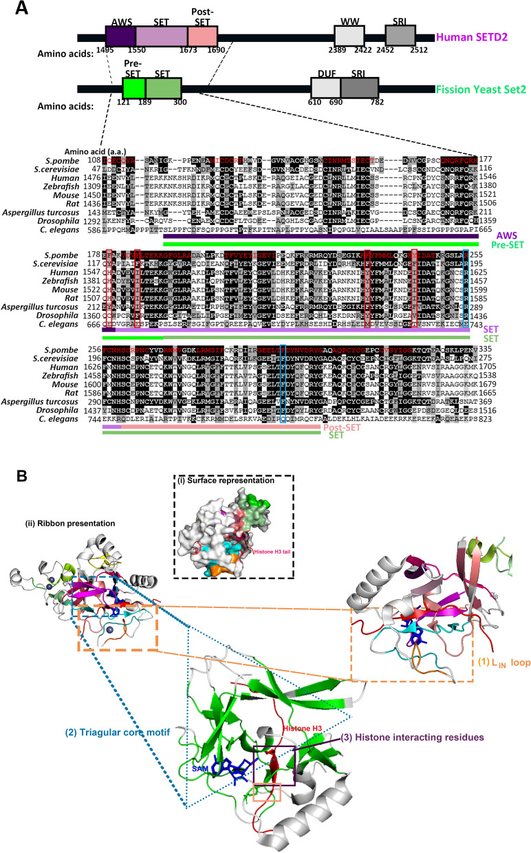
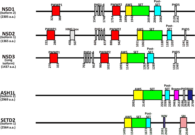
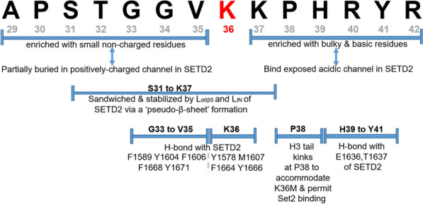
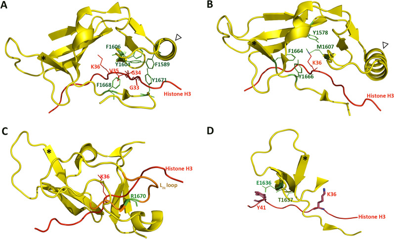
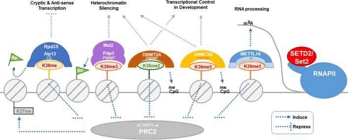
The methylation of histone H3 at lysine 36 (H3K36me) is essential for maintaining genomic stability. Indeed, this methylation mark is essential for proper transcription, recombination, and DNA damage response. Loss- and gain-of-function mutations in H3K36 methyltransferases are closely linked to human developmental disorders and various cancers. Structural analyses suggest that nucleosomal components such as the linker DNA and a hydrophobic patch constituted by histone H2A and H3 are likely determinants of H3K36 methylation in addition to the histone H3 tail, which encompasses H3K36 and the catalytic SET domain. Interaction of H3K36 methyltransferases with the nucleosome collaborates with regulation of their auto-inhibitory changes fine-tunes the precision of H3K36me in mediating dimethylation by NSD2 and NSD3 as well as trimethylation by Set2/SETD2. The identification of specific structural features and various cis-acting factors that bind to different forms of H3K36me, particularly the di-(H3K36me2) and tri-(H3K36me3) methylated forms of H3K36, have highlighted the intricacy of H3K36me functional significance. Here, we consolidate these findings and offer structural insight to the regulation of H3K36me2 to H3K36me3 conversion. We also discuss the mechanisms that underlie the cooperation between H3K36me and other chromatin modifications (in particular, H3K27me3, H3 acetylation, DNA methylation and N6-methyladenosine in RNAs) in the physiological regulation of the epigenomic functions of chromatin.

Keywords: ASH1L; H3K36; Methylation; NSD2; NSD3; SETD2; Set2.


Conflict of interest statement

The authors declare no conflict of interests.

Figures

Fig. 1
Fig. 1
Set2/SETD2 is generally conserved among fungi and metazoans. A Multiple sequence alignment (MSA) of Set2 homologs across fungi and metazoan species. MEGAX software and NCBI MSA viewer 1.13.1 [230] were used to generate an alignment of the primary amino acid sequences of Set2/SETD2 homologs from fission yeast, budding yeast, fungi Aspergillus turcosus, and metazoan species including human, rat, mouse, zebrafish, fruitfly, and nematode worm C. elegans. 35% of the human SETD2 amino acid sequence is conserved in fission yeast Set2 , even for sequences beyond the catalytic SET domain. 53% of the amino sequence in the catalytic SET and post-SET domains is identical in fission yeast Set2 and human SETD2. Residues that are generally conserved across species are indicated in red. Residues that are identical or similar in polarity across species are, respectively, highlighted in black or grey. Conserved “decision-making” residues that regulate the degree of methylation are circled in blue. Non-conserved aromatic residues that make contact with histone tails and possibly participate in methylation regulation are circled in red. Domain structure of fission yeast Set2 and human SETD2 are also shown to indicate the relative amino acid positions of the MSA sequences in the SETD2 homologues. B Conserved motifs in human SETD2 SET-domain. i Surface representation: sections of conserved motifs are highlighted (green, teal, cyan, and orange). Pymol visualization is derived from the crystallized structure database in the Protein Data Bank, entry 5V21 [74]. ii Projection of the conserved sequence of yeast Set2 and human SETD2. The SET domain crystalized structure highlights (1) a regulatory LIN loop, (2) a triangular core motif separated from the catalytic site, and (3) histone-interacting residues (refer Figs. 3 and 4). Conserved residues in the triple β-sheet in the triangular core of the SETD2 SET domain (green) endow the SET domain with its recognizable triangular shape, which maintains the structure of the domain. Conserved LIN-loop (teal, cyan and orange) and α8 (short, white α-helix region) in the closed conformation secure the histone tail in position for methylation.
Fig. 2
Fig. 2
Schematic diagram detailing the domain structure of common isoforms of NSD methyltransferases, ASH1L and SETD2. Numbers listed vertically refer to the relative amino acid positions of the domains in the different methyltransferases
Fig. 3
Fig. 3
SETD2 interaction with K36 and its flanking residues of Histone H3.3 residue from A29-R42. Note that the summary is based on results derived from histone H3 peptides with M36K substitution, which stabilizes protein interaction [75, 145]
Fig. 4
Fig. 4
Conserved histone-interacting residues in the SET domain of SETD2. Asterisk (*) and white arrow in the figure, respectively, label the position of SETD2 β15 beta sheet and α6 helix to depict relative orientation of SETD2 crystalized structure in different sub-figures. A F1589, Y1604, F1606, F1668, and Y1671 (green) interact with G33-V35 (dark red) of the histone H3 peptide (red) via hydrogen bonding. B Key catalytic residues Tyr-1578, Met-1607, Phe-1664 and Tyr-1666 (green) of the SETD2 SET domain (yellow) surround the K36/M36 residues (dark red) of the histone H3 peptide (red). C Key auto-inhibitory residue R1670 (green) of the LIN loop (brown) in SETD2 SET domain (yellow) is in proximity to K36 (dark red) on histone H3 peptide (red). D E1636 and T1637 (green) interact with Y41 (dark red) of the histone H3 peptide (red) via hydrogen bonding. Pymol visualization is derived from the crystalized structure database in the Protein Data Bank entry 5V22 [160].
Fig. 5
Fig. 5
Proposed crosstalk between di- and tri-methylated H3K36 and other modifications on histones, DNA, and RNA. Abbreviations: K36me: di- and/or tri-methylated H3K36, K36me2: dimethylated H3K36, K36me3: tri-methylated H3K36, ac: histone acetylation, CD: chromodomain, PWWP: proline-tryptophan-tryptophan-proline motif, m6A: N6-methyladenosine RNA modification, RNAPII: RNA polymerase II, meCpG: methylated cytosine in CpG motif on DNA. Rpd3S-K36me interaction is observed in human, budding yeast and fission yeast; Mst2-Pdp3-K36me interaction is reported solely from fission yeast system thus far, while interactions of DNMTs and MTTL14-K36me are studies from human

Similar articles

Cited by

References

    1. Luger K, Dechassa ML, Tremethick DJ. New insights into nucleosome and chromatin structure: an ordered state or a disordered affair? Nat Rev Mol Cell Biol. 2012;13:436–447. doi: 10.1038/nrm3382. - DOI - PMC - PubMed
    1. Luger K, Mäder AW, Richmond RK, Sargent DF, Richmond TJ. Crystal structure of the nucleosome core particle at 2.8 Å resolution. Nature. 1997;389:251–260. doi: 10.1038/38444. - DOI - PubMed
    1. Woodcock CL, Skoultchi AI, Fan Y. Role of linker histone in chromatin structure and function: H1 stoichiometry and nucleosome repeat length. Chromosome Res. 2006;14:17–25. doi: 10.1007/s10577-005-1024-3. - DOI - PubMed
    1. Bednar J, Garcia-Saez I, Boopathi R, Cutter AR, Papai G, Reymer A, et al. Structure and dynamics of a 197 bp nucleosome in complex with linker histone H1. Mol Cell. 2017;66:384–397.e8. doi: 10.1016/j.molcel.2017.04.012. - DOI - PMC - PubMed
    1. Healton SE, Pinto HD, Mishra LN, Hamilton GA, Wheat JC, Swist-Rosowska K, et al. H1 linker histones silence repetitive elements by promoting both histone H3K9 methylation and chromatin compaction. Proc Natl Acad Sci. 2020;117:14251–14258. doi: 10.1073/pnas.1920725117. - DOI - PMC - PubMed

Publication types

LinkOut - more resources