

Estrogenic regulation of reproduction and energy homeostasis by a triumvirate of hypothalamic arcuate neurons
- PMID: 35581942
- PMCID: PMC10228899
- DOI: 10.1111/jne.13145
Estrogenic regulation of reproduction and energy homeostasis by a triumvirate of hypothalamic arcuate neurons
Abstract
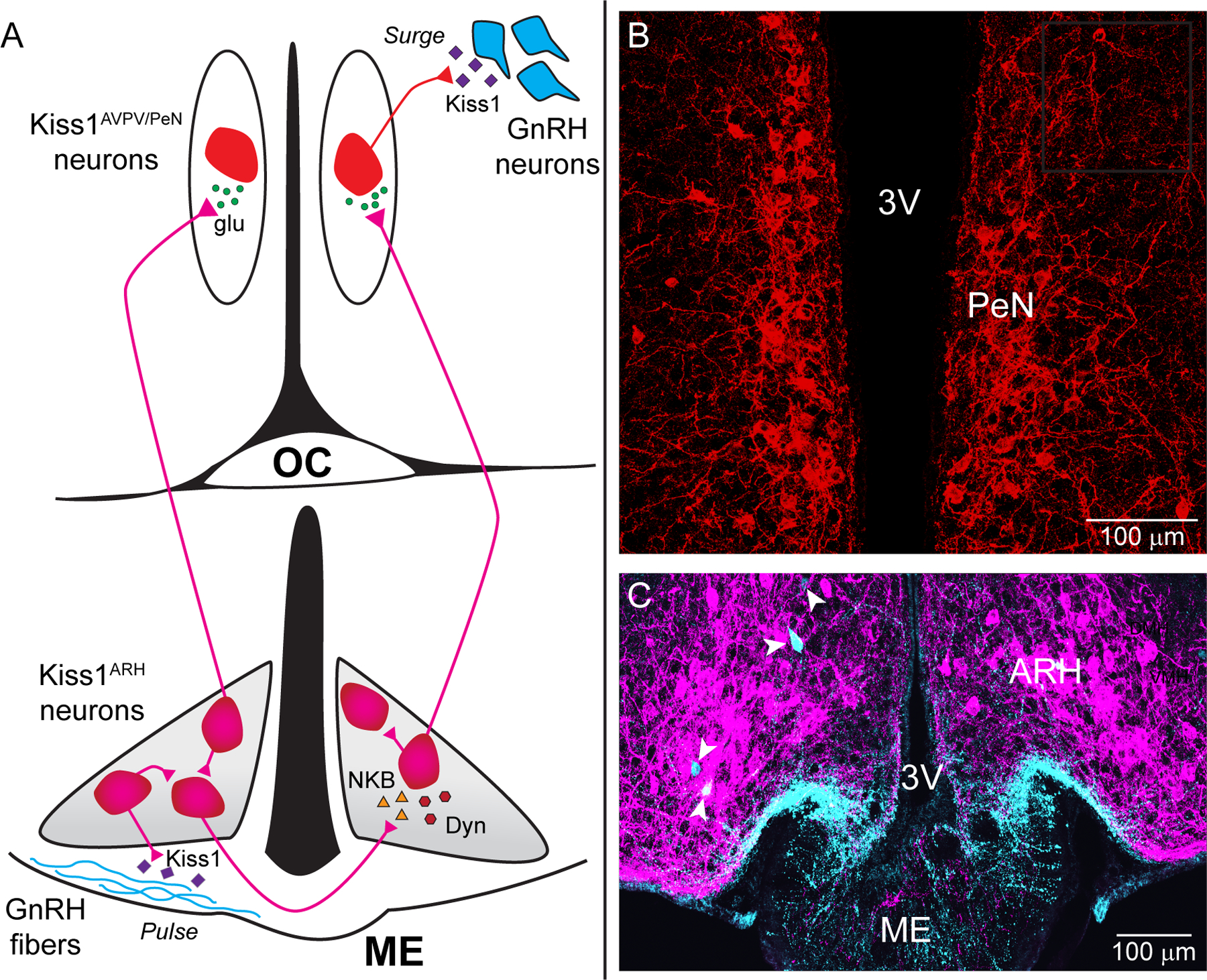
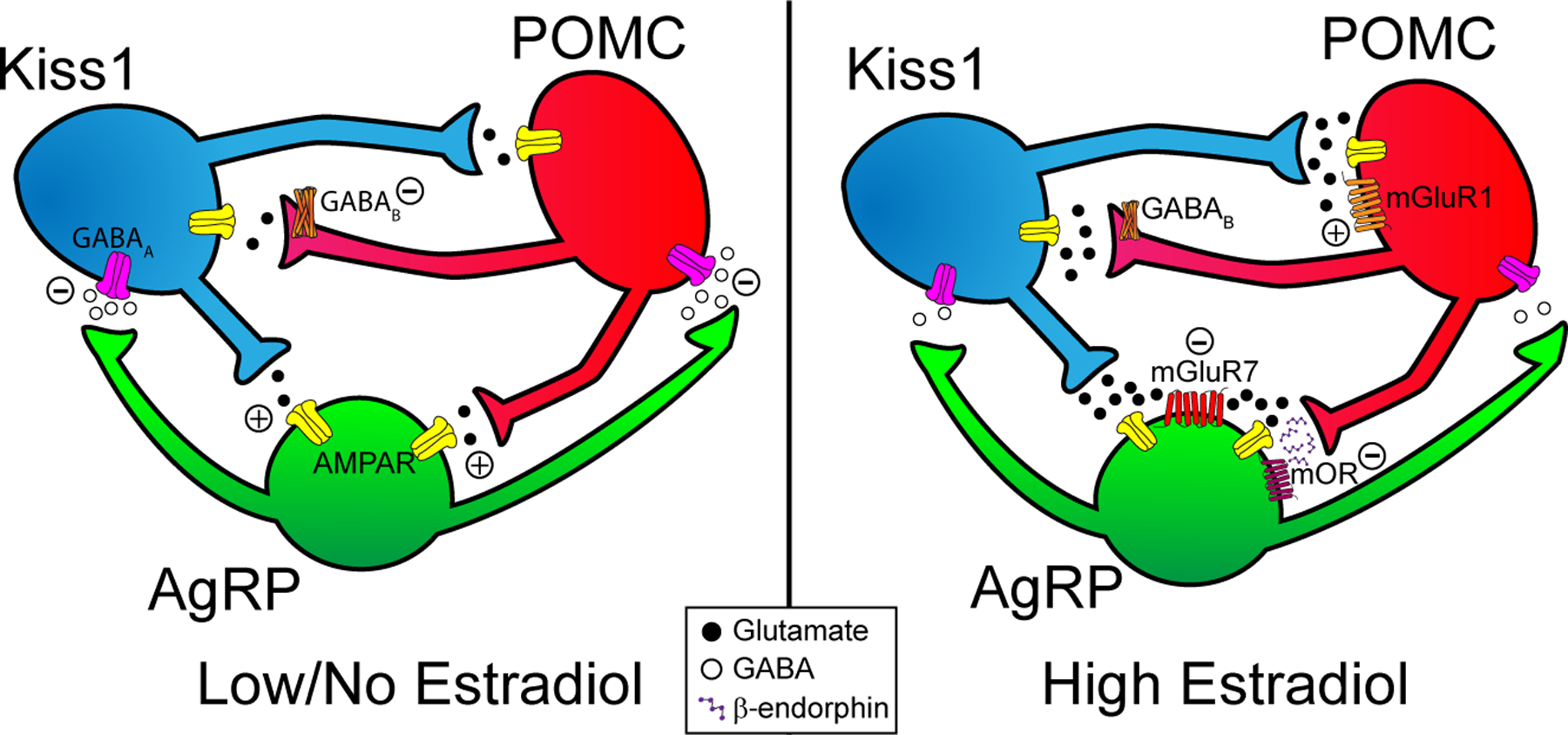
Pregnancy is energetically demanding and therefore, by necessity, reproduction and energy balance are inextricably linked. With insufficient or excessive energy stores a female is liable to suffer complications during pregnancy or produce unhealthy offspring. Gonadotropin-releasing hormone neurons are responsible for initiating both the pulsatile and subsequent surge release of luteinizing hormone to control ovulation. Meticulous work has identified two hypothalamic populations of kisspeptin (Kiss1) neurons that are critical for this pattern of release. The involvement of the hypothalamus is unsurprising because its quintessential function is to couple the endocrine and nervous systems, coordinating energy balance and reproduction. Estrogens, more specifically 17β-estradiol (E2 ), orchestrate the activity of a triumvirate of hypothalamic neurons within the arcuate nucleus (ARH) that govern the physiological underpinnings of these behavioral dynamics. Arising from a common progenitor pool, these cells differentiate into ARH kisspeptin, pro-opiomelanocortin (POMC), and agouti related peptide/neuropeptide Y (AgRP) neurons. Although the excitability of all these subpopulations is subject to genomic and rapid estrogenic regulation, Kiss1 neurons are the most sensitive, reflecting their integral function in female fertility. Based on the premise that E2 coordinates autonomic functions around reproduction, we review recent findings on how Kiss1 neurons interact with gonadotropin-releasing hormone, AgRP and POMC neurons, as well as how the rapid membrane-initiated and intracellular signaling cascades activated by E2 in these neurons are critical for control of homeostatic functions supporting reproduction. In particular, we highlight how Kiss1 and POMC neurons conspire to inhibit AgRP neurons and diminish food motivation in service of reproductive success.
Keywords: agouti-related peptide; hypothalamus; kisspeptin neurons; neuropeptide Y; pro-opiomelanocortin.
© 2022 British Society for Neuroendocrinology.
Figures
Similar articles
-
Membrane and nuclear initiated estrogenic regulation of homeostasis.Steroids. 2021 Apr;168:108428. doi: 10.1016/j.steroids.2019.108428. Epub 2019 Jun 20. Steroids. 2021. PMID: 31229508 Free PMC article. Review.
-
Arcuate Kisspeptin Neurons Coordinate Reproductive Activities with Metabolism.Semin Reprod Med. 2019 May;37(3):131-140. doi: 10.1055/s-0039-3400251. Epub 2019 Dec 23. Semin Reprod Med. 2019. PMID: 31869841 Free PMC article. Review.
-
Deletion of Stim1 in Hypothalamic Arcuate Nucleus Kiss1 Neurons Potentiates Synchronous GCaMP Activity and Protects against Diet-Induced Obesity.J Neurosci. 2021 Nov 24;41(47):9688-9701. doi: 10.1523/JNEUROSCI.0622-21.2021. Epub 2021 Oct 15. J Neurosci. 2021. PMID: 34654752 Free PMC article.
-
AgRP to Kiss1 neuron signaling links nutritional state and fertility.Proc Natl Acad Sci U S A. 2017 Feb 28;114(9):2413-2418. doi: 10.1073/pnas.1621065114. Epub 2017 Feb 14. Proc Natl Acad Sci U S A. 2017. PMID: 28196880 Free PMC article.
-
Optogenetic Stimulation of Arcuate Nucleus Kiss1 Neurons Reveals a Steroid-Dependent Glutamatergic Input to POMC and AgRP Neurons in Male Mice.Mol Endocrinol. 2016 Jun;30(6):630-44. doi: 10.1210/me.2016-1026. Epub 2016 Apr 19. Mol Endocrinol. 2016. PMID: 27093227 Free PMC article.
Cited by
-
Neuroactive steroids in the neuroendocrine control of food intake, metabolism, and reproduction.Endocrine. 2024 Sep;85(3):1050-1057. doi: 10.1007/s12020-024-03755-x. Epub 2024 Apr 18. Endocrine. 2024. PMID: 38635064 Review.
-
Comparative Hypothalamic Transcriptome Analysis Reveals Crucial mRNAs, lncRNAs, and circRNAs Affecting Litter Size in Goats.Genes (Basel). 2023 Feb 9;14(2):444. doi: 10.3390/genes14020444. Genes (Basel). 2023. PMID: 36833370 Free PMC article.
-
Obesity Alters POMC and Kisspeptin Neuron Cross Talk Leading to Reduced Luteinizing Hormone in Male Mice.J Neurosci. 2024 Jul 10;44(28):e0222242024. doi: 10.1523/JNEUROSCI.0222-24.2024. J Neurosci. 2024. PMID: 38744532 Free PMC article.
-
Hypothalamic Estrogen Receptor α Is Essential for Female Marmoset Sexual Behavior Without Protecting From Obesity.J Endocr Soc. 2025 Feb 5;9(3):bvaf012. doi: 10.1210/jendso/bvaf012. eCollection 2025 Feb 4. J Endocr Soc. 2025. PMID: 39911518 Free PMC article.
-
High sucrose consumption decouples intrinsic and synaptic excitability of AgRP neurons without altering body weight.Int J Obes (Lond). 2023 Mar;47(3):224-235. doi: 10.1038/s41366-023-01265-w. Epub 2023 Feb 1. Int J Obes (Lond). 2023. PMID: 36725979 Free PMC article.
References
-
- King JC, Tobet SA, Snavely FL, Arimura AA. The LHRH system in normal and neonatally androgenized female rats. Peptides. 1980; 185–100.
-
- Lehman MN, Robinson JE, Karsch FJ, Silverman AJ. Immunocytochemical localization of luteinizing hormone-releasing hormone(LHRH) pathways in the sheep brain during anestrous and the mid-luteal phase of the estrous cycle. The Journal of Comparative Neurology. 1986; 24419–35. - PubMed
-
- Stincic TL, Qiu J, Connors AM, Kelly MJ, Rønnekleiv OK. Arcuate and Preoptic Kisspeptin neurons exhibit differential projections to hypothalamic nuclei and exert opposite postsynaptic effects on hypothalamic paraventricular and dorsomedial nuclei in the female mouse. eneuro. 2021ENEURO.0093–21.2021. - PMC - PubMed
-
- Silverman AJ. Distribution of luteinizing hormone-releasing hormone (LH-RH) in the guinea pig brain. Endocrinology. 1976; 9930–41. - PubMed
-
- Silverman AJ, Antunes JL, Abrams GM, Nilaver G, Thau R, Robinson JA, Ferin M, Krey LC. The luteinizing hormone-releasing hormone pathway in rhesus (Macaca mulatta) and pigtailed (Macaca nemestrina) monkeys: New observations on thick, unembedded sections. The Journal of Comparative Neurology. 1982; 211309–17. - PubMed
Publication types
MeSH terms
Substances
Grants and funding
LinkOut - more resources
Full Text Sources
Miscellaneous
