

Noninvasive imaging of hepatic dysfunction: A state-of-the-art review
- PMID: 35581963
- PMCID: PMC9048786
- DOI: 10.3748/wjg.v28.i16.1625
Noninvasive imaging of hepatic dysfunction: A state-of-the-art review
Abstract
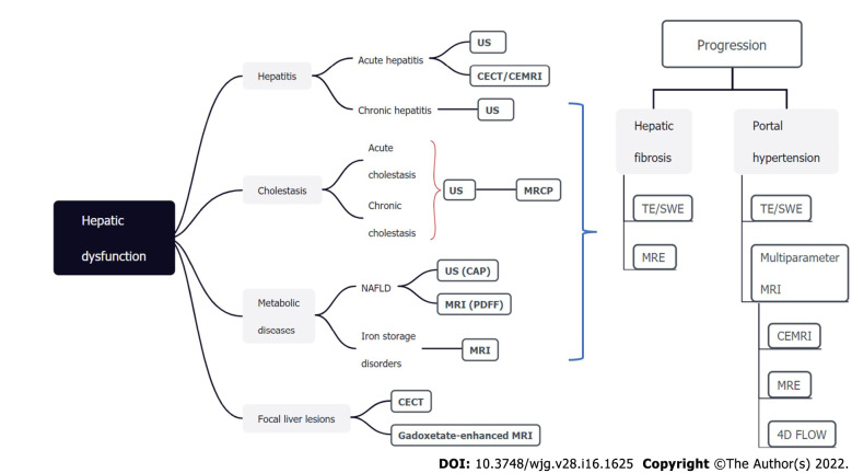
Hepatic dysfunction represents a wide spectrum of pathological changes, which can be frequently found in hepatitis, cholestasis, metabolic diseases, and focal liver lesions. As hepatic dysfunction is often clinically silent until advanced stages, there remains an unmet need to identify affected patients at early stages to enable individualized intervention which can improve prognosis. Passive liver function tests include biochemical parameters and clinical grading systems (e.g., the Child-Pugh score and Model for End-Stage Liver Disease score). Despite widely used and readily available, these approaches provide indirect and limited information regarding hepatic function. Dynamic quantitative tests of liver function are based on clearance capacity tests such as the indocyanine green (ICG) clearance test. However, controversial results have been reported for the ICG clearance test in relation with clinical outcome and the accuracy is easily affected by various factors. Imaging techniques, including ultrasound, computed tomography, and magnetic resonance imaging, allow morphological and functional assessment of the entire hepatobiliary system, hence demonstrating great potential in evaluating hepatic dysfunction noninvasively. In this article, we provide a state-of-the-art summary of noninvasive imaging modalities for hepatic dysfunction assessment along the pathophysiological track, with special emphasis on the imaging modality comparison and selection for each clinical scenario.
Keywords: Computed tomography; Hepatic dysfunction; Magnetic resonance imaging; Ultrasound.
©The Author(s) 2022. Published by Baishideng Publishing Group Inc. All rights reserved.
Conflict of interest statement
Conflict-of-interest statement: The authors have no conflict of interests related to this study.
Figures
Similar articles
-
Effectiveness comparison of indocyanine green retention test with the cirrhotic severity scoring in evaluating the pathological severity of liver cirrhosis in patients with hepatocellular carcinoma and Child-Pugh grade A liver function.World J Surg Oncol. 2020 Apr 23;18(1):79. doi: 10.1186/s12957-020-01854-3. World J Surg Oncol. 2020. PMID: 32326968 Free PMC article.
-
Plasma disappearance rate of indocyanine green in liver dysfunction.Transplant Proc. 2006 Apr;38(3):801-2. doi: 10.1016/j.transproceed.2006.01.049. Transplant Proc. 2006. PMID: 16647475
-
Comparative study of indocyanine green-R15, Child-Pugh score, and model for end-stage liver disease score for prediction of hepatic encephalopathy after transjugular intrahepatic portosystemic shunt.World J Gastroenterol. 2021 Feb 7;27(5):416-427. doi: 10.3748/wjg.v27.i5.416. World J Gastroenterol. 2021. PMID: 33584073 Free PMC article.
-
Current Modalities for the Assessment of Future Remnant Liver Function.Visc Med. 2017 Dec;33(6):442-448. doi: 10.1159/000480385. Epub 2017 Nov 30. Visc Med. 2017. PMID: 29344518 Free PMC article. Review.
-
Predictive factors of short term outcome after liver transplantation: A review.World J Gastroenterol. 2016 Jul 14;22(26):5936-49. doi: 10.3748/wjg.v22.i26.5936. World J Gastroenterol. 2016. PMID: 27468188 Free PMC article. Review.
Cited by
-
Hepatocytic Ballooning in Non-alcoholic Steatohepatitis: Bridging the Knowledge Gap and Charting Future Avenues.Cureus. 2023 Sep 25;15(9):e45884. doi: 10.7759/cureus.45884. eCollection 2023 Sep. Cureus. 2023. PMID: 37885505 Free PMC article. Review.
References
-
- Agrawal S, Dhiman RK, Limdi JK. Evaluation of abnormal liver function tests. Postgrad Med J. 2016;92:223–234. - PubMed
-
- Dillon JF, Miller MH, Robinson EM, Hapca A, Rezaeihemami M, Weatherburn C, McIntyre PG, Bartlett B, Donnan PT, Boyd KA, Dow E. Intelligent liver function testing (iLFT): A trial of automated diagnosis and staging of liver disease in primary care. J Hepatol. 2019;71:699–706. - PubMed
-
- Dhiman RK, Agrawal S, Gupta T, Duseja A, Chawla Y. Chronic Liver Failure-Sequential Organ Failure Assessment is better than the Asia-Pacific Association for the Study of Liver criteria for defining acute-on-chronic liver failure and predicting outcome. World J Gastroenterol. 2014;20:14934–14941. - PMC - PubMed
-
- Bajaj JS, O’Leary JG, Reddy KR, Wong F, Biggins SW, Patton H, Fallon MB, Garcia-Tsao G, Maliakkal B, Malik R, Subramanian RM, Thacker LR, Kamath PS North American Consortium For The Study Of End-Stage Liver Disease (NACSELD) Survival in infection-related acute-on-chronic liver failure is defined by extrahepatic organ failures. Hepatology. 2014;60:250–256. - PMC - PubMed
Publication types
MeSH terms
Substances
LinkOut - more resources
Full Text Sources
Medical
Research Materials
