Reevaluation of the expanded indications in undifferentiated early gastric cancer for endoscopic submucosal dissection
- PMID: 35582127
- PMCID: PMC9048457
- DOI: 10.3748/wjg.v28.i15.1548
Reevaluation of the expanded indications in undifferentiated early gastric cancer for endoscopic submucosal dissection
Abstract
Background: Although the criteria for the indication of endoscopic submucosal dissection (ESD) for undifferentiated early gastric cancer (UD-EGC) have been recently proposed, accumulating reports on the non-negligible rate of lymph node metastasis (LNM) after indicated ESD raise questions on the reliability of the current criteria.
Aim: To investigate the prevalence and risk factors of LNM in UD-EGC cases meeting the expanded indication for ESD.
Methods: We retrospectively reviewed 4780 UD-EGC cases that underwent surgical resection between January 2008 and February 2019 at Asan Medical Center, a tertiary university hospital in Korea. To identify the risk factors of LNM of UD-EGC meeting the expanded criteria for ESD, we performed a case-control study by matching the cases with LNM to those without at a ratio of 1:4. We reviewed the clinical, endoscopic, and histologic features of the cases to identify features with a significant difference according to the presence of LNM. Univariate and multivariate logistic regression analyses were performed to estimate the odds ratios (ORs).
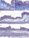
Results: Of the 4780 UD-EGC cases, 1240 (25.9%) were identified to meet the expanded indication for ESD. Of the 1240 cases, 14 (1.1%) cases had LNM. Among the various clinical, endoscopic, and histopathological features that were evaluated, mixed histology (tumors consisting of 10%-90% of signet ring cells) had a marginally significant association (P = 0.059) with the risk of LNM. Moreover, diffuse blurring of the muscularis mucosae (MM) underneath the tumorous epithelium, a previously unrecognized histologic feature, had a significant association with the absence of LNM (P = 0.028). Multivariate logistic regression analysis showed that the blurring of MM was the only explanatory variable significantly associated with a reduced risk of LNM (OR: 0.12, 95%CI: 0.02-0.95; P = 0.045).
Conclusion: The risk of LNM is higher than expected when using the current expanded indication for UD-EGC. Histological evaluation could provide useful clues for reducing the risk of LNM.
Keywords: Endoscopic submucosal dissection; Gastric cancer; Lymph node metastasis; Undifferentiated carcinoma.
©The Author(s) 2022. Published by Baishideng Publishing Group Inc. All rights reserved.
Conflict of interest statement
Conflict-of-interest statement: All authors have no conflict of interest related to the study.
Figures


References
-
- Gotoda T, Yanagisawa A, Sasako M, Ono H, Nakanishi Y, Shimoda T, Kato Y. Incidence of lymph node metastasis from early gastric cancer: estimation with a large number of cases at two large centers. Gastric Cancer. 2000;3:219–225. - PubMed
-
- Kang HJ, Kim DH, Jeon TY, Lee SH, Shin N, Chae SH, Kim GH, Song GA, Srivastava A, Park DY, Lauwers GY. Lymph node metastasis from intestinal-type early gastric cancer: experience in a single institution and reassessment of the extended criteria for endoscopic submucosal dissection. Gastrointest Endosc. 2010;72:508–515. - PubMed
Publication types
MeSH terms
LinkOut - more resources
Full Text Sources
Medical
Miscellaneous

