

Non-destructive monitoring of 3D cell cultures: new technologies and applications
- PMID: 35582620
- PMCID: PMC9107788
- DOI: 10.7717/peerj.13338
Non-destructive monitoring of 3D cell cultures: new technologies and applications
Abstract
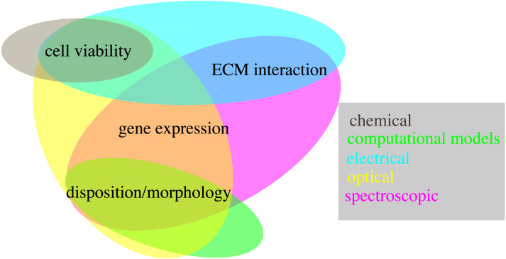
3D cell cultures are becoming the new standard for cell-based in vitro research, due to their higher transferrability toward in vivo biology. The lack of established techniques for the non-destructive quantification of relevant variables, however, constitutes a major barrier to the adoption of these technologies, as it increases the resources needed for the experimentation and reduces its accuracy. In this review, we aim at addressing this limitation by providing an overview of different non-destructive approaches for the evaluation of biological features commonly quantified in a number of studies and applications. In this regard, we will cover cell viability, gene expression, population distribution, cell morphology and interactions between the cells and the environment. This analysis is expected to promote the use of the showcased technologies, together with the further development of these and other monitoring methods for 3D cell cultures. Overall, an extensive technology shift is required, in order for monolayer cultures to be superseded, but the potential benefit derived from an increased accuracy of in vitro studies, justifies the effort and the investment.
Keywords: 3D cell culture; Non-destructive technology; in-vitro quantitative analysis.
©2022 Cortesi and Giordano.
Conflict of interest statement
The authors declare there are no competing interests.
Figures


Similar articles
-
Direct lysis of 3D cell cultures for RT-qPCR gene expression quantification.Sci Rep. 2023 Jan 27;13(1):1520. doi: 10.1038/s41598-023-28844-1. Sci Rep. 2023. PMID: 36707637 Free PMC article.
-
Volumetric imaging of human mesenchymal stem cells (hMSCs) for non-destructive quantification of 3D cell culture growth.PLoS One. 2023 Mar 28;18(3):e0282298. doi: 10.1371/journal.pone.0282298. eCollection 2023. PLoS One. 2023. PMID: 36976801 Free PMC article.
-
Cell Viability Assays in Three-Dimensional Hydrogels: A Comparative Study of Accuracy.Tissue Eng Part C Methods. 2021 Jul;27(7):401-410. doi: 10.1089/ten.TEC.2021.0060. Tissue Eng Part C Methods. 2021. PMID: 34082602 Free PMC article.
-
3D in vitro corneal models: A review of current technologies.Exp Eye Res. 2020 Nov;200:108213. doi: 10.1016/j.exer.2020.108213. Epub 2020 Sep 3. Exp Eye Res. 2020. PMID: 32890484 Free PMC article. Review.
-
Devices and techniques used to obtain and analyze three-dimensional cell cultures.Biotechnol Prog. 2021 May;37(3):e3126. doi: 10.1002/btpr.3126. Epub 2021 Jan 29. Biotechnol Prog. 2021. PMID: 33460298 Review.
Cited by
-
Driving cell response through deep learning, a study in simulated 3D cell cultures.Heliyon. 2024 Apr 23;10(9):e29395. doi: 10.1016/j.heliyon.2024.e29395. eCollection 2024 May 15. Heliyon. 2024. PMID: 38699000 Free PMC article.
-
New approach methodologies (NAMs) for the in vitro assessment of cleaning products for respiratory irritation: workshop report.Front Toxicol. 2024 Oct 8;6:1431790. doi: 10.3389/ftox.2024.1431790. eCollection 2024. Front Toxicol. 2024. PMID: 39439531 Free PMC article. Review.
-
Design approaches for 3D cell culture and 3D bioprinting platforms.Biophys Rev (Melville). 2024 May 16;5(2):021304. doi: 10.1063/5.0188268. eCollection 2024 Jun. Biophys Rev (Melville). 2024. PMID: 38765221 Free PMC article. Review.
-
Improvements in Maturity and Stability of 3D iPSC-Derived Hepatocyte-like Cell Cultures.Cells. 2023 Sep 27;12(19):2368. doi: 10.3390/cells12192368. Cells. 2023. PMID: 37830581 Free PMC article.
-
A comparative analysis of 2D and 3D experimental data for the identification of the parameters of computational models.Sci Rep. 2023 Sep 22;13(1):15769. doi: 10.1038/s41598-023-42486-3. Sci Rep. 2023. PMID: 37737283 Free PMC article.
References
-
- Abbas M, Moradi F, Hu W, Regudo KL, Osborne M, Pettipas J, Atallah DS, Hachem R, Ott-Peron N, Stuart JA. Vertebrate cell culture as an experimental approach–limitations and solutions. Comparative Biochemistry and Physiology Part B: Biochemistry and Molecular Biology. 2021;254:110570. doi: 10.1016/j.cbpb.2021.110570. - DOI - PubMed
-
- Accardo A, Blatché M-C, Courson R, Loubinoux I, Vieu C, Malaquin L. Two-photon lithography and microscopy of 3D hydrogel scaffolds for neuronal cell growth. Biomedical Physics & Engineering Express. 2018;4(2):027009. doi: 10.1088/2057-1976/aaab93. - DOI
-
- Ahlfeld T, Cubo-Mateo N, Cometta S, Guduric V, Vater C, Bernhardt A, Akkineni AR, Lode A, Gelinsky M. A novel plasma-based bioink stimulates cell proliferation and differentiation in bioprinted, mineralized constructs. ACS Applied Materials & Interfaces. 2020;12(11):12557–12572. doi: 10.1021/acsami.0c00710. - DOI - PubMed
Publication types
MeSH terms
LinkOut - more resources
Full Text Sources
