. 2022 May;13(55):3261-3265.
doi: 10.1016/j.med.2022.05.009. Epub 2022 May 12.

[Diagnostic protocol for febrile lung infiltrates during the COVID-19 pandemic]

[Article in Spanish]
Affiliations

[Diagnostic protocol for febrile lung infiltrates during the COVID-19 pandemic]

[Article in Spanish]
R Henche Sánchez et al. Medicine (Madr). 2022 May.

Abstract

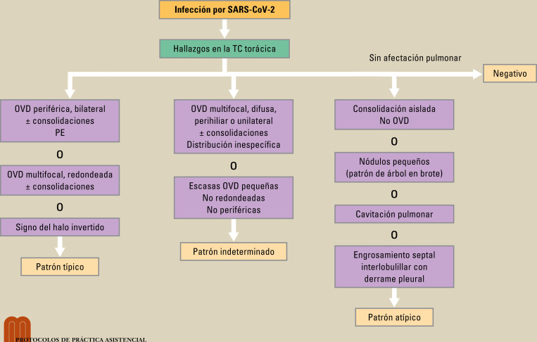
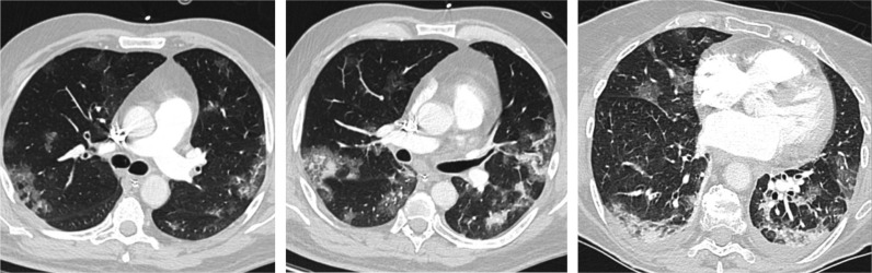
Chest x-ray and computed tomography (CT) scans are important pillars for the diagnosis of lung involvement in COVID-19. The radiological image is typically characterized by peripheral, bilateral ground glass opacities (GGO), mainly located in the lower lobes. The limited sensitivity and specificity of these imaging techniques and possible atypical morphological or topographical presentations make it necessary to always rule out other infectious and non-infectious diseases. Therefore, it is fundamental to consider the patient's clinical and analytical data and the epidemiological circumstances.

La radiografía de tórax y la tomografía computarizada (TC) son pilares importantes para el diagnóstico de la afectación pulmonar en la COVID-19, con una imagen radiológica caracterizada típicamente por opacidades en vidrio deslustrado (OVD) periféricas, bilaterales y localizadas principalmente en lóbulos inferiores. La sensibilidad y la especificidad limitada de estas técnicas de imagen y las posibles presentaciones morfológicas o topográficas atípicas obligan a descartar siempre otras patologías tanto infecciosas como no infecciosas, para lo que es fundamental considerar los datos clínicos y analíticos del paciente y las circunstancias epidemiológicas.

Keywords: COVID-19; Chest x-ray; Computed tomography; Differential diagnosis.


Figures

Fig. 1
Fig. 1
Hallazgos radiológicos en la tomografía computarizada (TC) torácica en la COVID-19. ?Modificada de Farias LPG, et al. OVD: opacidades en vidrio deslustrado; PE: patrón en empedrado; TC: tomografía computarizada.
Fig. 2
Fig. 2
Diferente grado de afectación pulmonar típica en la COVID-19. Opacidades en vidrio deslustrado de distribución predominantemente periférica o más difusa, con patrón en empedrado.

Similar articles

References

    1. fx4 Rubin G.D., Haramati L.B., Kane J.P., Schluger N.W., Yim J.-J., Anderson D.J., et al. The role of chest imaging in patient management during the COVID-19 pandemic: A multinational consensus statement from the Fleischner Society. Radiology. 2020;296:172–180. - PMC - PubMed
    1. fx4 Martínez-Chamorro E., Díez Tascón A., Ibáñez-Sanz L., Ossaba-Vélez S., Borruel-Nacenta S. Manejo diagnóstico inicial del paciente con COVID-19. Radiología. 2021;63(1):56–73. - PMC - PubMed
    1. fx4 Wong H.Y.F., Lam H.Y.S., Fong A.H.-T., Leung S.T., Chin T.W.-Y., Lo C.S.Y., et al. Frequency and distribution of chest radiographic findings in COVID-19 positive patients. Radiology. 2020;296(2):E72–E78. - PMC - PubMed
    1. fx4 Bai H.X., Hsieh B., Xiong Z., Halsey K., Choi J.W., Tran T.M.L., et al. Performance of radiologists in differentiating COVID-19 from viral pneumonia at chest CT. Radiology. 2020;296(2):E46–E54. - PMC - PubMed
    1. fx4 fx1 Farias L.P.G., Strabelli D.G., Fonseca E.K.U.N., Loureiro B.M.C., Nomura C.H., Sawamura M.V.Y. Thoracic tomographic manifestations in symptomatic respiratory patients with COVID-19. Radiol Bras. 2020;53(4):255–261. - PMC - PubMed

Publication types

LinkOut - more resources