. 2022 Jun;161(6):478-491.
doi: 10.1111/jnc.15647. Epub 2022 May 30.

Signaling pathways involved in NMDA-induced suppression of M-channels in corticotropin-releasing hormone neurons in central amygdala

Affiliations

Signaling pathways involved in NMDA-induced suppression of M-channels in corticotropin-releasing hormone neurons in central amygdala

Hua Zhang et al. J Neurochem. 2022 Jun.

Abstract

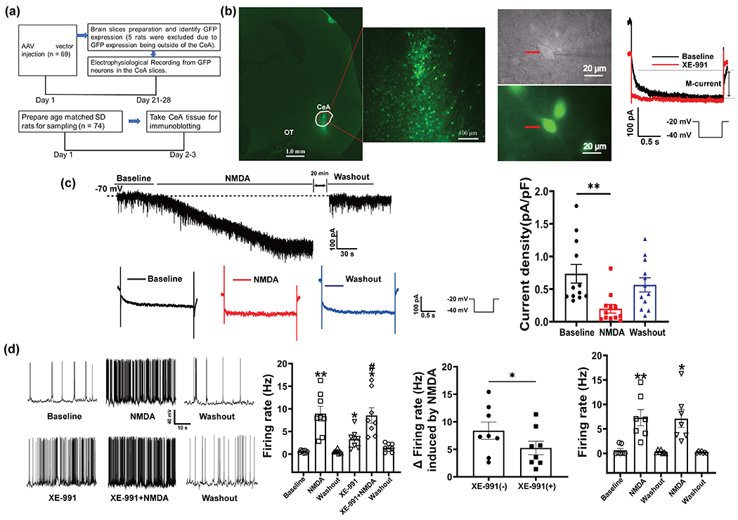
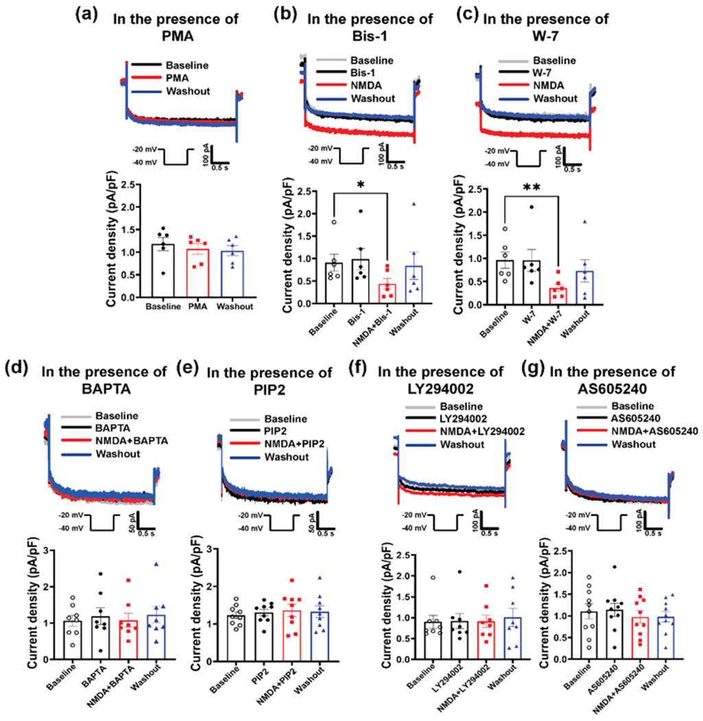
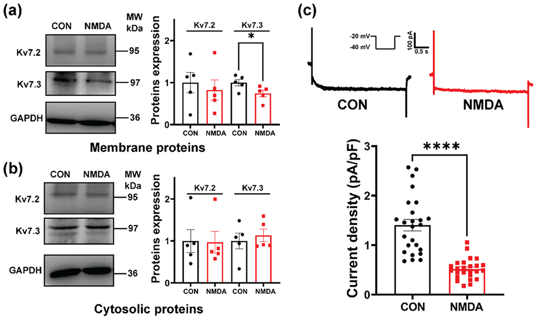
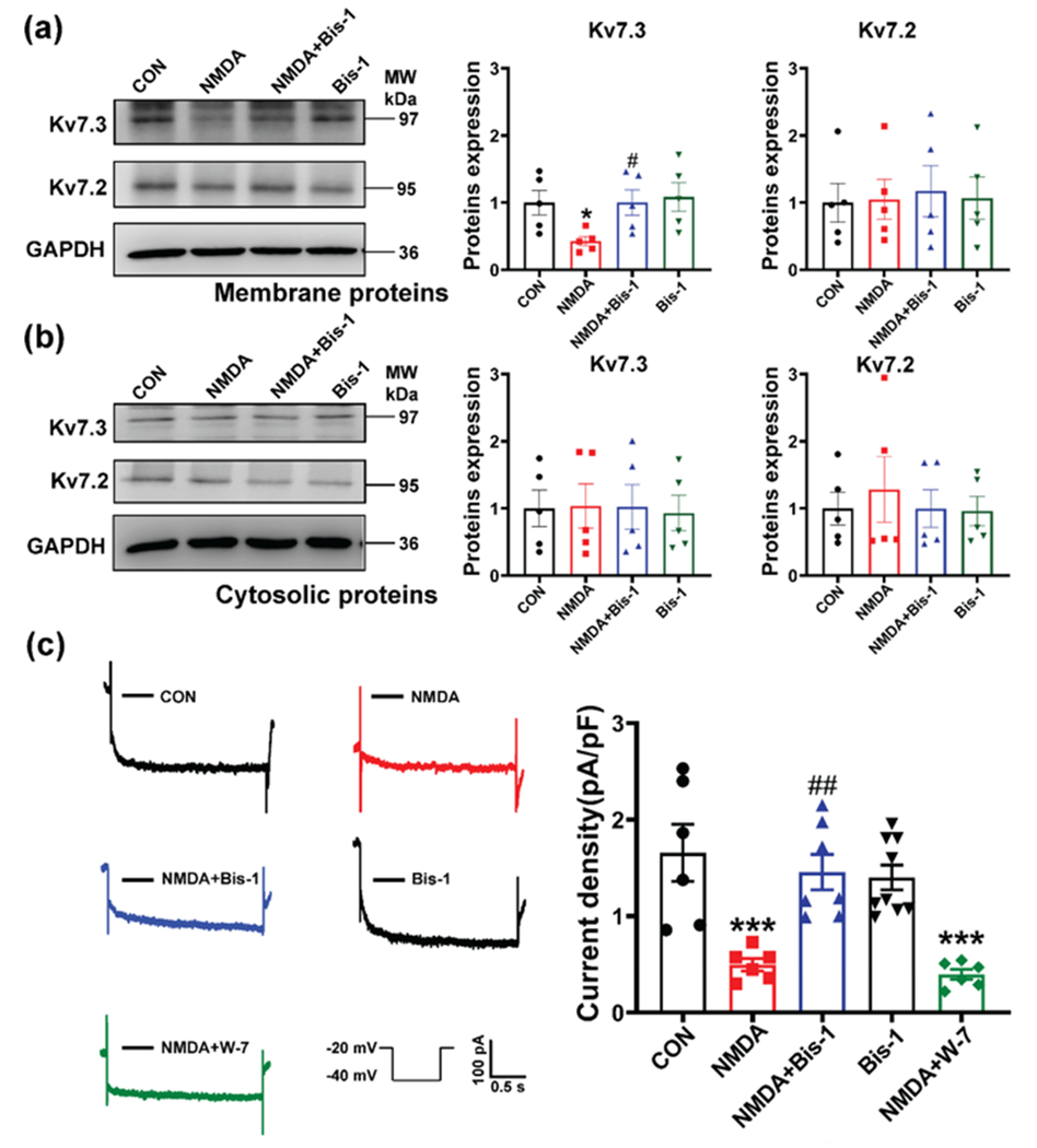
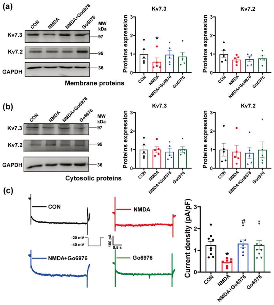
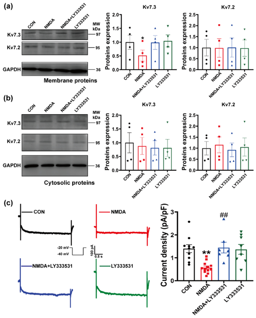
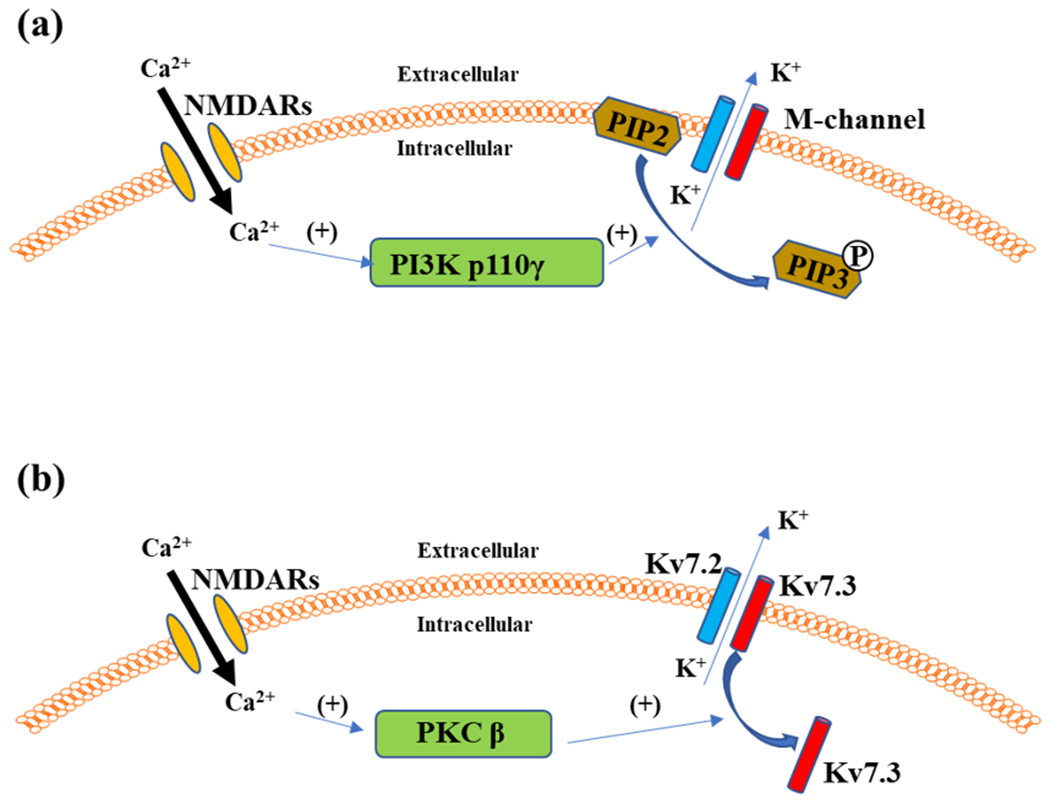
Glutamate N-methyl-d-aspartate (NMDA) receptors (NMDARs) and Kv7/M channels are importantly involved in regulating neuronal activity involved in various physiological and pathological functions. Corticotropin-releasing hormone (CRH)-expressing neurons in the central nucleus of the amygdala (CeA) critically mediate autonomic response during stress. However, the interaction between NMDA receptors and Kv7/M channels in the CRHCeA neurons remains unclear. In this study, we identified rat CRHCeA neurons through the expression of an AAV viral vector-mediated enhanced green fluorescent protein (eGFP) driven by the rat CRH promoter. M-currents carried by Kv7/M channels were recorded using the whole-cell patch-clamp approach in eGFP-tagged CRHCeA neurons in brain slices. Acute exposure to NMDA significantly reduced M-currents recorded from the CRHCeA neurons. NMDA-induced suppression of M-currents was eliminated by chelating intracellular Ca2+ , supplying phosphatidylinositol 4,5-bisphosphate (PIP2) intracellularly, or blocking phosphoinositide3-kinase (PI3K). In contrast, inhibiting protein kinase C (PKC) or calmodulin did not alter NMDA-induced suppression of M-currents. Sustained exposure of NMDA decreased Kv7.3 membrane protein levels and suppressed M-currents, while the Kv7.2 expression levels remained unaltered. Pre-treatment of brain slices with PKC inhibitors alleviated the decreases in Kv7.3 and reduction of M-currents in CRHCeA neurons induced by NMDA. PKC inhibitors did not alter Kv7.2 and Kv7.3 membrane protein levels and M-currents in CRHCeA neurons. These data suggest that transient activation of NMDARs suppresses M-currents through the Ca2+ -dependent PI3K-PIP2 signaling pathway. In contrast, sustained activation of NMDARs reduces Kv7.3 protein expression and suppresses M-currents through a PKC-dependent pathway.

Keywords: Corticotropin-releasing hormone (CRH)-expressing neurons; Kv7/M channel; NMDA receptor; amygdala; protein kinase C.


Conflict of interest statement

Conflict(s) of Interest/Disclosure(s)

The authors declare no competing financial interests.

Figures

Figure 1.
Figure 1.. Transient activation of NMDARs inhibited M-currents in CRHCeA neurons.
(a) Graphical timeline of the experimental procedure. (b) Left panel: representative images show the microinjection site and labeled neurons in CeA. Middle panel: microphotographs show the eGFP-tagged neurons in the CeA under light and florescent illuminant. Right panel: M-currents were triggered by voltage pulses from −20 to −40 mV and then returned to −20 mV. M-currents were defined as XE-991-sensitive currents. (c) Representative traces and summary data show M-currents before and after bath application of 20 μM NMDA (n =12 neurons from 6 rats, p = 0.0012, Kruskal-Wallis test). (d) Representative traces and summary data show firing activity recoded from CRHCeA neurons in the presence of NMDA (n = 8 neurons from 3 rats, p = 0.0087) and NMDA plus XE-991 (n = 8 neurons from 3 rats, p = 0.015, one-way ANOVA). The upper and lower traces were obtained from the same neuron. Please note that NMDA-induced increase in firing activity was attenuated in the presence of XE-991 (n = 8 neurons from 3 rats, p = 0.011 vs. XE-991(−), two-tailed paired t-test). * p < 0.05, ** p < 0.01 versus basal value, # p < 0.05 vs. XE-991 group.
Figure 2.
Figure 2.. Intracellular pathways involved in reducing M-currents induced by transient activation of NMDARs.
(a) Representative traces and summary data show that bath application of PKC activator PMA did not change M-currents in CRHCeA neurons (n = 6 neurons from 3 rats, p = 0.75, one-way ANOVA). (b) and (c) Representative traces and summary data show M-currents recorded with the inclusion of PKC inhibitor Bis-1 (b, n = 6 neurons from 3 rats, p = 0.024, Kruskal-Wallis test) or calmodulin inhibitor W-7 (c, n = 6 neurons from 3 rats, p = 0.0386, Kruskal-Wallis test) in the internal solution. Please note that Bis-1 and W-7 did not affect NMDA-induced inhibition of M-currents. (d) Representative traces and summary data show that the inclusion of BAPTA in the internal solution abolished NMDA-induced inhibition of M-currents in CRHCeA neurons in rats (n = 8 neurons from 3 rats, p = 0.7634, Kruskal-Wallis test). (e) Representative traces and summary data depict M-currents recorded with the inclusion of synthetic PIP2 in the internal solution. Application of NMDA did not change M-currents recorded with PIP2 in the internal solution in CRHCeA neurons (n = 9 neurons from 3 rats, p = 0.9569, one-way ANOVA). (f) and (g) Representative traces and summary data show inclusion of LY294002, a PI3K inhibitor (f, n = 8 neurons from 3 rats, p = 0.9827, Kruskal-Wallis test), or AS605240, a PI3Kγ inhibitor (g, n = 10 neurons from 3 rats, p = 0.6328, one-way ANOVA), in the internal solution eliminated NMDA-induced suppression of M-currents in CRHCeA neurons. * p < 0.05, ** p < 0.01 versus basal value.
Figure 3.
Figure 3.. Sustained activation of NMDARs decreased Kv7.3 membrane protein level and reduced M-currents in CRHCeA neurons.
(a) and (b) Effects of sustained application of NMDA on the membrane and cytosolic protein levels of Kv7.2 and Kv7.3 in CeA tissues from vehicle-treated and NMDA-treated brain slices. NMDA incubation for 40 mins significantly reduced Kv7.3 membrane expression level (n =5 samples, p = 0.0474, t(8) = 2.34, two-tailed unpaired t-test) but did not change Kv7.2 membrane level (n =5 samples, p = 0.6215, t(8) = 0.5135, two-tailed unpaired t-test) and cytosolic protein levels for Kv7.3 (n =5 samples, p > 0.99, two-tailed Mann-Whitney test) and Kv7.2 (n =5 samples, p = 0.8413, two-tailed Mann-Whitney test). Each sample was from tissue in one rat. (c) Representative traces and summary data depict M-currents recorded in CRHCeA neurons incubated with vehicle (n = 25 neurons from 9 rats) or NMDA (n = 25 neurons from 9 rats) for 40 mins. M-current density was significantly decreased in CRHCeA neurons treated with NMDA for 40 mins (p < 0.0001, t(48) = 7.211, two-tailed unpaired t-test). * p < 0.05, **** p < 0.0001 versus CON value.
Figure 4.
Figure 4.. PKC inhibitor alleviated Kv7 channel suppression induced by sustained activation of NMDARs.
(a) and (b) Effect of long-lasting incubation (40 mins) of NMDA on the membrane (a) and cytosolic (b) proteins of Kv7.2 and Kv7.3 in the presence and absence of PKC inhibitor Bis-1. Each sample was from tissue in 1 rat. n = 5 samples in each group. (c) Raw tracings and summary data show M-currents recorded from CRHCeA neurons in brain slices treated by vehicle, NMDA, Bis-1, NMDA plus Bis-1, or NMDA plus W-7 for 40 mins (n= 6-9 neurons from 3 rats in each group). Please note that incubation of NMDA significantly reduced Kv7.3 (but not Kv7.2) membrane protein level (p = 0.0461, one-way ANOVA) and M-currents (p = 0.0006, one-way ANOVA) in CRHCeA neurons. Treatment with Bis-1 alleviated the reduction of Kv7.3 membrane protein level (p = 0.0424 vs. NMDA incubation, one-way ANOVA) and M-currents (p = 0.0036 vs. NMDA incubation, one-way ANOVA) in CRHCeA neurons. W-7 did not change the reduced M-currents. * p < 0.05, *** p < 0.001 versus CON value; # p < 0.05, ## p < 0.01 versus NMDA incubation group.
Figure 5.
Figure 5.. PKC isoforms were involved in reducing Kv7.3 channel membrane expression levels and M-currents induced by sustained activation of NMDARs.
(a) and (b) Effects of long-lasting NMDA incubation (40 mins) on the membrane (a) and cytosolic (b) proteins of Kv7.2 and Kv7.3 in the presence of Go6976 (inhibitor for PKCα and PKCβ). Co-incubation of Go6976 with NMDA eliminated the decrease in Kv7.3 membrane protein levels in CeA tissue induced by NMDA incubation (p = 0.0156, one-way ANOVA). Each sample was from 1 rat and 5 samples in each group. (c) M-currents were recorded in CRHCeA neurons in brain slices treated by NMDA for 40 mins in the presence or absence of Go6976 (n= 7-9 neurons from 3 rats in each group). Treatment with Go6976 alone did not change M-current density in CRHCeA neurons, but co-incubation of Go6976 with NMDA (p = 0.0127 vs. NMDA incubation, one-way ANOVA) eliminated NMDA-induced reduction of M-currents (p = 0.0154, one-way ANOVA). * p < 0.05 versus CON value; # p < 0.05 versus NMDA incubation group.
Figure 6.
Figure 6.. PKCβ inhibitor alleviated Kv7 channel suppression induced by long-lasting activation of NMDARs.
(a) and (b) Effect of long-lasting incubation (40 mins) of NMDA on the membrane (a) and cytosolic (b) proteins of Kv7.2 and Kv7.3 in the presence and absence of PKCβ inhibitor, LY333531. Each sample was from tissue in 1 rat. n = 4 samples in each group. NMDA incubation decreased Kv7.3 membrane protein levels in CeA tissue (p = 0.0416, one-way ANOVA), whereas co-incubation of the tissue with NMDA plus LY333531 did not change Kv7.3 membrane protein levels (p = 0.9986, one-way ANOVA). (c) Representative recordings and summary data show M-currents recorded in CRHCeA neurons in brain slice treated by NMDA for 40 mins in the presence or absence of LY333531, (n=7-12 neurons from 3 rats in each group). NMDA significantly reduced M-currents (p = 0.0026, one-way ANOVA), and this effect was abolished by co-incubation of LY333531 (p = 0.0044 vs. NMDA incubation, one-way ANOVA). * p < 0.05, ** p < 0.01 versus CON value; ## p < 0.01 versus NMDA incubation group.
Figure 7.
Figure 7.
The schematic diagram depicts two distinct mechanisms involved in suppressing Kv7 channel activity by NMDARs. (a) Transient activation of NMDARs induces Ca2+ influx to trigger PI3Kp110γ-mediated phosphorylation of PIP2 to PIP3, a process that causes PIP2 reduction and subsequent Kv7 channel suppression. (b) Sustained activation of NMDARs decreases Kv7.3 membrane protein levels and suppresses Kv7 channel activity through Ca2+-dependent activation of the PKCβ isoform.

Similar articles

Cited by

References

    1. Alaimo A and Villarroel A (2018) Calmodulin: a multitasking protein in Kv7.2 potassium channel functions. Biomolecules 8. - PMC - PubMed
    1. Andersen MN, Olesen SP and Rasmussen HB (2011) Kv7.1 surface expression is regulated by epithelial cell polarization. Am J Physiol Cell Physiol 300, C814–824. - PubMed
    1. Brennan-Minnella AM, Shen Y, El-Benna J and Swanson RA (2013) Phosphoinositide 3-kinase couples NMDA receptors to superoxide release in excitotoxic neuronal death. Cell Death Dis 4, e580. - PMC - PubMed
    1. Brown DA and Passmore GM (2009) Neural KCNQ (Kv7) channels. Br J Pharmacol 156, 1185–1195. - PMC - PubMed
    1. Cullen PJ (2003) Calcium signalling: the ups and downs of protein kinase C. Curr Biol 13, R699–701. - PubMed

Publication types

MeSH terms

Substances