. 2022 Jun 17;479(11):1149-1164.
doi: 10.1042/BCJ20210770.

Characterization of uridine-cytidine kinase like-1 nucleoside kinase activity and its role in tumor growth

Affiliations

Characterization of uridine-cytidine kinase like-1 nucleoside kinase activity and its role in tumor growth

Emily C Matchett et al. Biochem J. .

Abstract

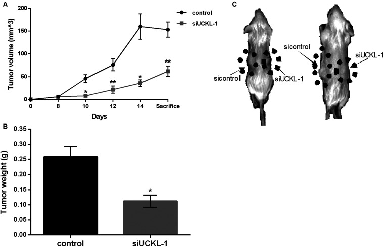
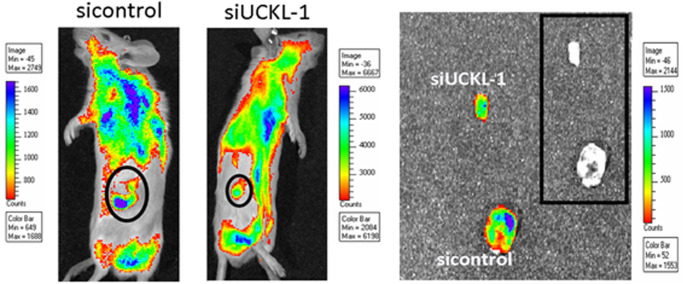
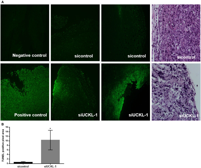
Uridine-cytidine kinase like-1 (UCKL-1) is a largely uncharacterized protein with high sequence similarity to other uridine-cytidine kinases (UCKs). UCKs play an important role in the pyrimidine salvage pathway, catalyzing the phosphorylation of uridine and cytidine to UMP and CMP, respectively. Only two human UCKs have been identified, UCK1 and UCK2. Previous studies have shown both enzymes phosphorylate uridine and cytidine using ATP as the phosphate donor. No studies have evaluated the kinase potential of UCKL-1. We cloned and purified UCKL-1 and found that it successfully phosphorylated uridine and cytidine using ATP as the phosphate donor. The catalytic efficiency (calculated as kcat/KM) was 1.2 × 10⁴ s-1, M-1 for uridine and 0.7 × 10⁴ s-1, M-1 for cytidine. Our lab has previously shown that UCKL-1 is up-regulated in tumor cells, providing protection against natural killer (NK) cell killing activity. We utilized small interfering RNA (siRNA) to down-regulate UCKL-1 in vitro and in vivo to determine the effect of UCKL-1 on tumor growth and metastasis. The down-regulation of UCKL-1 in YAC-1 lymphoma cells in vitro resulted in decreased cell counts and increased apoptotic activity. Down-regulation of UCKL-1 in K562 leukemia cells in vivo led to decreased primary tumor growth and less tumor cell dissemination and metastasis. These results identify UCKL-1 as a bona fide pyrimidine kinase with the therapeutic potential to be a target for tumor growth inhibition and for diminishing or preventing metastasis.

Keywords: cancer; metastasis; natural killer; nucleoside kinase; uridine kinase.


Conflict of interest statement

The authors declare that there are no competing interests associated with the manuscript.

Figures

Figure 1.
Figure 1.. Alignment of human UCKL-1, UCK1, and UCK2.
Human UCKL-1 (UniprotKB Q9NWZ5), UCK1 (Q9HA47) and UCK2 (Q9BZX2) were aligned using the Clustal program. Identical amino acids among the three proteins are highlighted in yellow. Similar amino acids between UCKL1 and either UCK1 or UCK2 are depicted in red. The boxed amino acids represent the ATP binding site. The section of UCKL-1 highlighted in blue has 41% similarity to human UPRT (Q96BW1).
Figure 2.
Figure 2.. Purification of bacterially expressed His-tagged UCKL-1 and UCK2.
(A) Coomassie blue stained gel of 100 ng of purified UCKL-1 (lane 1) and UCK2 (lane 2). Single band detected for each: UCKL-1 at 80 kDa and UCK2 at 48 kDa. (B) Western blot analysis with His antibody confirmed purity and isolation of His-tagged UCKL-1 (lane 1) and UCK2 (lane 2). (C) Analysis with UCKL-1 (B-11) antibody (lane 1) showed no cross-reactivity with UCK2 (lane 2). (D) Analysis with UCK2 antibody (lane 2) showed no cross-reactivity with UCKL-1 (lane 1). Blots represent 1 of 3 experiments with identical results.
Figure 3.
Figure 3.. Pyrimidine kinase activity of UCKL-1 and UCK2.
Purified UCKL-1 (500 ng) and UCK2 (1 ng) were dispensed into 384-well plates and mixed with a substrate/ATP buffer. Final concentration of ATP was 100 µM. ADP formation was measured at various substrate concentrations. (A) UCKL-1 with uridine as substrate; (B) UCK2 with uridine as substrate; (C) UCKL-1 with cytidine as substrate; (D) UCK2 with cytidine as substrate. Kinase reactions were measured at 0, 60, 120, and 180 min. Luminescence at each time point was converted to the amount of ADP formed during the kinase reaction using an ATP-to-ADP conversion curve. ADP formation was plotted versus time. Each data point represents the mean ± SD of 3–5 replicates.
Figure 4.
Figure 4.. Immunoblot analysis of UCKL-1, UCK1, and UCK2 expression in various tumor cell lines.
(A) Whole-cell lysates were prepared and UCKL-1, UCK1, and UCK2 were detected by immunoblotting. Actin was used as a loading control. (BD) Densitometric analysis of UCKL-1, UCK1, and UCK2 expression relative to actin for each cell line (mean ± SD, n = 3).
Figure 5.
Figure 5.. Down-regulation of UCKL-1 in YAC-1 cells induces apoptosis.
(A) YAC-1 cells were nucleofected with either siGFP control (siGFP) or siUCKL-1. Cell counts were significantly lower at 16, 20, and 24 h after nucleofection of siUCKL-1 compared with siGFP (mean ± SD, n = 5, * P ≤ 0.02, ** P ≤ 0.002, *** P ≤ 0.0002). UCKL-1 down-regulation was confirmed by real-time PCR 24 h after nucleofection. (B) YAC-1 cells nucleofected with siUCKL-1 had more caspase 3/7 activity than siGFP-treated cells (mean ± SD, n = 4, * P ≤ 0.02). Caspase activity was determined by a luminescent assay and UCKL-1 down-regulation confirmed by real-time PCR.
Figure 6.
Figure 6.. UCKL-1 down-regulation inhibits primary tumor growth in NSG mice.
(A) 106 K562-GFP cells suspended in Matrigel were injected into the right and left flanks of NSG mice. Once tumors were established (day 8), either 8 µg scrambled siRNA (sicontrol) or UCKL-1 siRNA (siUCKL-1) in 50 µl PBS was injected intratumorally. Tumors were measured and injected with siRNA every other day for one week. Significantly less tumor growth was seen in the siUCKL-1-injected tumors at day 10, 12, 14, and day of sacrifice (day 15) (mean ± SD, n = 18, * P ≤ 0.001, ** P ≤ 0.0005). (B) Subcutaneous tumors were extracted from NSG mice on day 15 and weighed. SiUCKL-1-injected tumors weighed significantly less than sicontrol-injected tumors (mean ± SD, n = 18, * P ≤ 0.001). (C) Representative images of NSG mice at sacrifice. The siUCKL-1-injected subcutaneous tumor on the right flanks of the mice were visibly smaller than the sicontrol-injected tumor on the left. Two representative mice are shown.
Figure 7.
Figure 7.. Diminished growth of siUCKL-1 treated tumors in vivo.
NSG mice were injected subcutaneously with K562-GFP tumor cells on both rear flanks. Tumors were injected with either scrambled (sicontrol) or UCKL-1 siRNA (siUCKL-1) every other day and measured. Representative IVIS images of NSG mice showed less growth of siUCKL-1-injected tumors. Tumors were visible by GFP both in the mouse (left) and after extraction (right). The levels of GFP were analyzed by IVIS software and quantitated by photon efficiency. There was 3-fold less GFP expression in the siUCKL-1 tumors compared with the sicontrol tumors (n = 18).
Figure 8.
Figure 8.. Less lung metastasis and fewer circulating tumor cells in mice receiving intratumoral injections of siUCKL-1.
(A) Lungs from tumor-bearing NSG mice were extracted and analyzed for GFP expression by quantitative RT-PCR. GFP expression correlates with the number of K562-GFP tumor cells present. The level of lung metastasis was significantly lower in mice injected with siUCKL-1 than in sicontrol-injected mice (mean ± SD, n = 34, ** P ≤ 0.01). (B) At time of sacrifice, 500 µl blood was removed from the heart. RNA was extracted and analyzed for GFP by quantitative RT-PCR. Mice bearing siUCKL-1-injected tumors had significantly fewer circulating tumor cells than mice bearing sicontrol-injected tumors (mean ± SD, n = 13, * P ≤ 0.04). No GFP expression was detected in mice without tumor present.
Figure 9.
Figure 9.. Higher levels of apoptosis in UCKL-1 down-regulated K562 tumors in vivo.
(A) Tumors isolated from NSG mice were fixed, paraffin embedded, sectioned, and subjected to fluorescent TUNEL staining, which visualizes nicks in the DNA and is reflective of apoptosis. A negative control and positive control (DNase treatment) were used for background and positive staining determination, respectively. TUNEL staining is depicted in green. Representative images are shown. siUCKL-1-injected tumors had higher levels of apoptosis than sicontrol-injected tumors. The two right panels are Hematoxylin and Eosin staining of the tumors. (B) Quantitative analyses of TUNEL staining. The TUNEL positive pixel area of six fields was analyzed for each tumor sample and normalized to negative control background staining. siUCKL-1-injected tumors had significantly more TUNEL fluorescent staining (mean ± SD, n = 8, * P ≤ 0.05).

References

    1. Qian, Y., Ding, Q., Li, Y., Zou, Z., Yan, B. and Ou, L. (2014) Phosphorylation of uridine and cytidine by uridine-cytidine kinase. J. Biotechnol. 188, 81–87 10.1016/j.jbiotec.2014.08.018 - DOI - PubMed
    1. Cheng, N., Payne, R.C. and Traut, T.W. (1986) Regulation of uridine kinase: evidence for a regulatory site. J. Biol. Chem. 261, 13006–13012 10.1016/S0021-9258(18)69262-2 - DOI - PubMed
    1. Van Rompay, A.R., Norda, A., Lindén, K., Johansson, M. and Karlsson, A. (2001) Phosphorylation of uridine and cytidine nucleoside analogs by two human uridine-cytidine kinases. Mol. Pharmacol. 59, 1181–1186 10.1124/mol.59.5.1181 - DOI - PubMed
    1. Suzuki, N.N., Koizumi, K., Fukushima, M., Matsuda, A. and Inagaki, F. (2004) Structural basis for the specificity, catalysis, and regulation of human uridine-cytidine kinase. Structure 12, 751–764 10.1016/j.str.2004.02.038 - DOI - PubMed
    1. Van Rompay, A.R., Johansson, M. and Karlsson, A. (1999) Phosphorylation of deoxycytidine analog monophosphates by UMP-CMP kinase: molecular characterization of the human enzyme. Mol. Pharmacol. 56, 562–569 10.1124/mol.56.3.562 - DOI - PubMed

Publication types