

Glioneuronal tumor with neuropil-like islands in the spinal cord: A case report and literature review
- PMID: 35583532
- PMCID: PMC9276349
- DOI: 10.1097/MD.0000000000029237
Glioneuronal tumor with neuropil-like islands in the spinal cord: A case report and literature review
Abstract
Rationale: Glioneuronal tumor with neuropil-like islands (GTNI) is a distinctive neoplasm located in the cerebrum. Moreover, spinal GTNI is extremely rare. Herein, we present a case of spinal GTNI and review the related literature.
Patient concerns: A 38-year-old Chinese woman presented to our hospital with a 6-month history of neck pain and a 1-month history of dizziness.
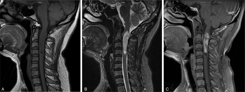
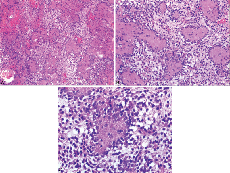
Diagnoses: Magnetic resonance imaging revealed a large intramedullary mass spanning the length of the spinal cord from C1 to C4. Microscopic and immunohistochemical examinations of the tumor tissue revealed findings typical of GTNI.
Interventions: The patient underwent C1 to C4 intraspinal gross tumor resection.
Outcomes: Follow-up results showed that the patient had no recurrence 6 months after tumor resection.
Lessons: GTNI in the spinal cord is a highly rare neoplasm with poor prognosis. Therefore, clinicians and pathologists should differentiate GTNI from other benign glioneuronal tumors, and long-term follow-up of patients with spinal GTNI is necessary.
Copyright © 2022 the Author(s). Published by Wolters Kluwer Health, Inc.
Conflict of interest statement
The authors have no funding and conflicts of interest to disclose.
Figures

References
-
- Teo JG, Gultekin SH, Bilsky M, Gutin P, Rosenblum MK. A distinctive glioneuronal tumor of the adult cerebrum with neuropil-like (including “rosetted”) islands: report of 4 cases. Am J Surg Pathol 1999;23:502–10. - PubMed
-
- Louis DN, Ohgaki H, Wiestler OD, et al. Pathology of Tumours of the Central Nervous System. 2007;Lyon, FR: IARC Press, 97–109.
-
- Harris BT, Horoupian DS. Spinal cord glioneuronal tumor with “rosetted” neuropil islands and meningeal dissemination: a case report. Acta Neuropathol 2000;100:575–9. - PubMed
-
- Ruppert B, Welsh CT, Hannah J, et al. Glioneuronal tumor with neuropil-like islands of the spinal cord with diffuse leptomeningeal neuraxis dissemination. J Neurooncol 2011;104:529–33. - PubMed
-
- Buccoliero AM, Castiglione F, Degl’innocenti DR, et al. Glioneuronal tumor with neuropil-like islands: clinical, morphologic, immunohistochemical, and molecular features of three pediatric cases. Pediatr Dev Pathol 2012;15:352–60. - PubMed
Publication types
MeSH terms
LinkOut - more resources
Full Text Sources
Medical
Miscellaneous
