

The Y plane is a reliable CT-based reference for glenoid component positioning in shoulder arthroplasty
- PMID: 35583701
- PMCID: PMC9117575
- DOI: 10.1186/s40634-022-00481-z
The Y plane is a reliable CT-based reference for glenoid component positioning in shoulder arthroplasty
Abstract
Purpose: To determine the reliability of anatomic references for mediolateral component positioning in shoulder arthroplasty.
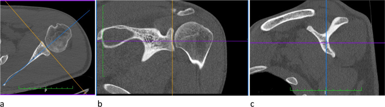
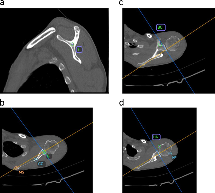
Materials and methods: The computed tomography scans of 86 shoulders free of arthritic or anatomic deformities were studied. Two surgeons independently digitized a series of points, including the intersection of the 3 bone branches of the scapular spine (Y), the center of the glenoid surface (G), the most medial point of the scapula (MS), the cortical convergence (CC) of the anterior and posterior margins of the glenoid, the base of the coracoid (BC), the anterior (HA) and posterior (HP) margins of the subchondral bone.
Results: The mean mediolateral distances between G and Y, BC, CC were respectively - 19.6 mm, - 1.5 mm, and - 36.8 mm. The consistency of anatomic landmarks was greatest for Y (standard deviation (SD) =2.3 mm; interquartile range (IQR) =3 mm), compared to BC (SD = 4.6 mm; IQR = 7 mm), and CC (SD = 6.6 mm; IQR = 8 mm). The repeatability of anatomic landmarks was excellent for all measurements. The mean ratios (relative to humeral head size) of distances between G and Y, BC, CC were respectively - 0.45, - 0.04, and - 0.85. The consistency of ratios was greatest for Y (SD = 0.05; IQR = 0.06), compared to BC (SD = 0.11; IQR = 0.14), and CC (SD = 0.13; IQR = 0.17). The repeatability of ratios was excellent for Y and BC, while it was good for CC.
Conclusions: The Y-plane is a reliable reference for glenoid component positioning in shoulder arthroplasty, with a consistent distance from the center of the glenoid surface, and could therefore be suitable for preoperative planning.
Study design: Level III, comparative anatomic study.
Keywords: Computed tomography; Lateralization; RSA; TSA; Y-point.
© 2022. The Author(s).
Conflict of interest statement
JMG declares receiving royalties and consultancy fees from Smith & Nephew. JK declares receiving royalties and consultancy fees from FH Orthopedics and consultancy from DePuy-Mitek (Johnson & Johnson), as well as consultancy fees from VIMS. FVR, MS and LN declare that they have no competing interests.
Figures
References
-
- Boileau P, Morin-Salvo N, Gauci MO, Seeto BL, Chalmers PN, Holzer N, Walch G. Angled BIO-RSA (bony-increased offset-reverse shoulder arthroplasty): a solution for the management of glenoid bone loss and erosion. J Shoulder Elb Surg. 2017;26(12):2133–2142. doi: 10.1016/j.jse.2017.05.024. - DOI - PubMed
LinkOut - more resources
Full Text Sources
Research Materials
Miscellaneous
