

Paradoxical venous air embolism detected with point-of-care ultrasound: a case report
- PMID: 35583704
- PMCID: PMC9116074
- DOI: 10.1186/s13089-022-00265-7
Paradoxical venous air embolism detected with point-of-care ultrasound: a case report
Abstract
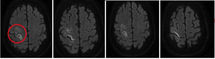
Venous air embolism (VAE) is an uncommon event consistent in the entrainment of air from any communication between the environment and the venous vasculature that could occur during central venous catheter (CVC) manipulation, and might trigger circulatory shock within minutes depending on the amount of air embolized. We present a case of a critical care patient who presented sudden clinical hemodynamic deterioration after the removal of central venous catheter. Hemodynamic evaluation with point-of-care ultrasound (POCUS) showed bubbles in both right and left heart cavities wherewith air embolism facilitated by heart septal defect was suspected. Therefore, the patient was reintubated, supported with vasopressors and a new CVC was inserted to proceed with air aspiration. Shortly after, the patient's hemodynamic status improved in terms of vital signs stabilization. 6 h after the event with optimal perfusion markers and diminished sedation, the patient showed left hemiparesis therefore a cerebral magnetic resonance (MRI) was also performed showing hyperintensity in the right precentral gyrus, so ischemic stroke without hemorrhagic transformation diagnosis was made, because of paradoxical embolism. This case report demonstrates the value of POCUS application as a diagnostic tool in the hemodynamically unstable patient.
Keywords: Air embolism; Case report; Point-of-care ultrasound; Stroke.
© 2022. The Author(s).
Conflict of interest statement
The authors confirm that there have been no financial or nonfinancial involvements in either authors that might raise the question of bias in the work reported or in the conclusions, implications, or opinions.
Figures
References
-
- Jain S, Kumar D, Chandra N, Kakkar M. Temporal insular glioma–rare case report for a venous air embolism. Ain-Shams J Anesthesiol. 2020;12(1):54. doi: 10.1186/s42077-020-00100-y. - DOI
LinkOut - more resources
Full Text Sources