. 2022 Oct;41(10):2848-2855.
doi: 10.1109/TMI.2022.3174561. Epub 2022 Sep 30.

Positronium Lifetime Image Reconstruction for TOF PET

Positronium Lifetime Image Reconstruction for TOF PET

Jinyi Qi et al. IEEE Trans Med Imaging. 2022 Oct.

Abstract

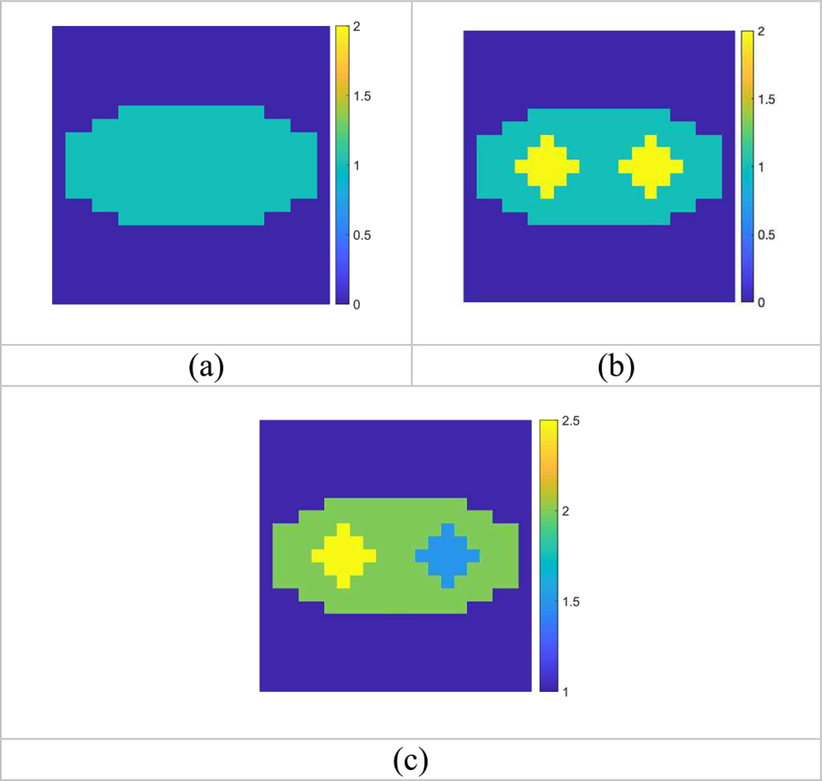
Positron emission tomography is widely used in clinical and preclinical applications. Positronium lifetime carries information about the tissue microenvironment where positrons are emitted, but such information has not been captured because of two technical challenges. One challenge is the low sensitivity in detecting triple coincidence events. This problem has been mitigated by the recent developments of PET scanners with long (1-2 m) axial field of view. The other challenge is the low spatial resolution of the positronium lifetime images formed by existing methods that is determined by the time-of-flight (TOF) resolution (200-500 ps) of existing PET scanners. This paper solves the second challenge by developing a new image reconstruction method to generate high-resolution positronium lifetime images using existing TOF PET. Simulation studies demonstrate that the proposed method can reconstruct positronium lifetime images at much better spatial resolution than the limit set by the TOF resolution of the PET scanner. The proposed method opens up the possibility of performing positronium lifetime imaging using existing TOF PET scanners. The lifetime information can be used to understand the tissue microenvironment in vivo which could facilitate the study of disease mechanism and selection of proper treatments.


Figures

Fig. 1.
Fig. 1.
The simulation phantom images. (a) True activity distribution (x) of phantom one. (b) True activity distribution (x) of phantom two. (c) True lifetime (in ns) distribution (λ−1) used in both phantoms.
Fig. 2.
Fig. 2.
Reconstructed activity and lifetime images (λ−1) of phantom one. (a) The activity image reconstructed by the ML-EM algorithm. (b) The lifetime image estimated by TOF backprojection. (c) Reconstructed lifetime image by the ML algorithm without regularization. (d) Reconstructed lifetime image by the PML algorithm with regularization. (e) The center horizontal profiles through the reconstructed lifetime images shown in (b)-(d) in comparison with the ground truth. For better visualization, lifetime values greater than 2.6 ns were set to 2.6 ns in the ML reconstruction.
Fig. 3.
Fig. 3.
The mean and standard deviation images of the lifetime images for phantom one. (a) the mean ML reconstruction; (b) the mean PML reconstruction (β = 10). (c) the standard deviation image of ML reconstruction; (d) the standard deviation of PML reconstruction (β = 10). S.d values above 0.3 ns were set to 0.3 ns.
Fig. 4.
Fig. 4.
The mean and standard deviation images of the lifetime images for phantom two. (a) the mean ML reconstruction; (b) the mean PML reconstruction (β = 10). (c) the standard deviation image of ML reconstruction; (d) the standard deviation of PML reconstruction (β = 10).
Fig. 5.
Fig. 5.
The objective function of the PML reconstruction with β = 10 as a function of iteration.

Similar articles

Cited by

References

    1. Beyer T et al., “A combined PET/CT scanner for clinical oncology,” Journal of nuclear medicine : official publication, Society of Nuclear Medicine, vol. 41, no. 8, pp. 1369–79, Aug 2000. [Online]. Available: http://www.ncbi.nlm.nih.gov/pubmed/10945530. - PubMed
    1. Gunn RN, Slifstein M, Searle GE, and Price JC, “Quantitative imaging of protein targets in the human brain with PET,” Physics in medicine and biology, Research Support, N.I.H., Extramural vol. 60, no. 22, pp. R363–411, Nov 21 2015, doi: 10.1088/0031-9155/60/22/R363. - DOI - PubMed
    1. Machac J, “Cardiac positron emission tomography imaging,” Seminars in nuclear medicine, Review vol. 35, no. 1, pp. 17–36, Jan 2005, doi: 10.1053/j.semnuclmed.2004.09.002. - DOI - PubMed
    1. Harpen MD, “Positronium: Review of symmetry, conserved quantities and decay for the radiological physicist,” Medical Physics, vol. 31, no. 1, pp. 57–61, 2004/January/01 2004, doi: 10.1118/1.1630494. - DOI - PubMed
    1. Shibuya K, Saito H, Nishikido F, Takahashi M, and Yamaya T, “Oxygen sensing ability of positronium atom for tumor hypoxia imaging,” Communications Physics, vol. 3, no. 1, p. 173, 2020/October/01 2020, doi: 10.1038/s42005-020-00440-z. - DOI

Publication types