. 2022 May 24;119(21):e2117349119.
doi: 10.1073/pnas.2117349119. Epub 2022 May 18.

Hydration-mediated G-protein-coupled receptor activation

Affiliations

Hydration-mediated G-protein-coupled receptor activation

Steven D E Fried et al. Proc Natl Acad Sci U S A. .

Abstract

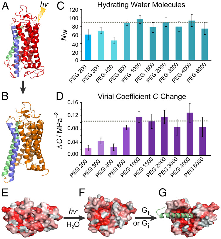
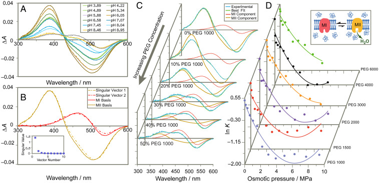
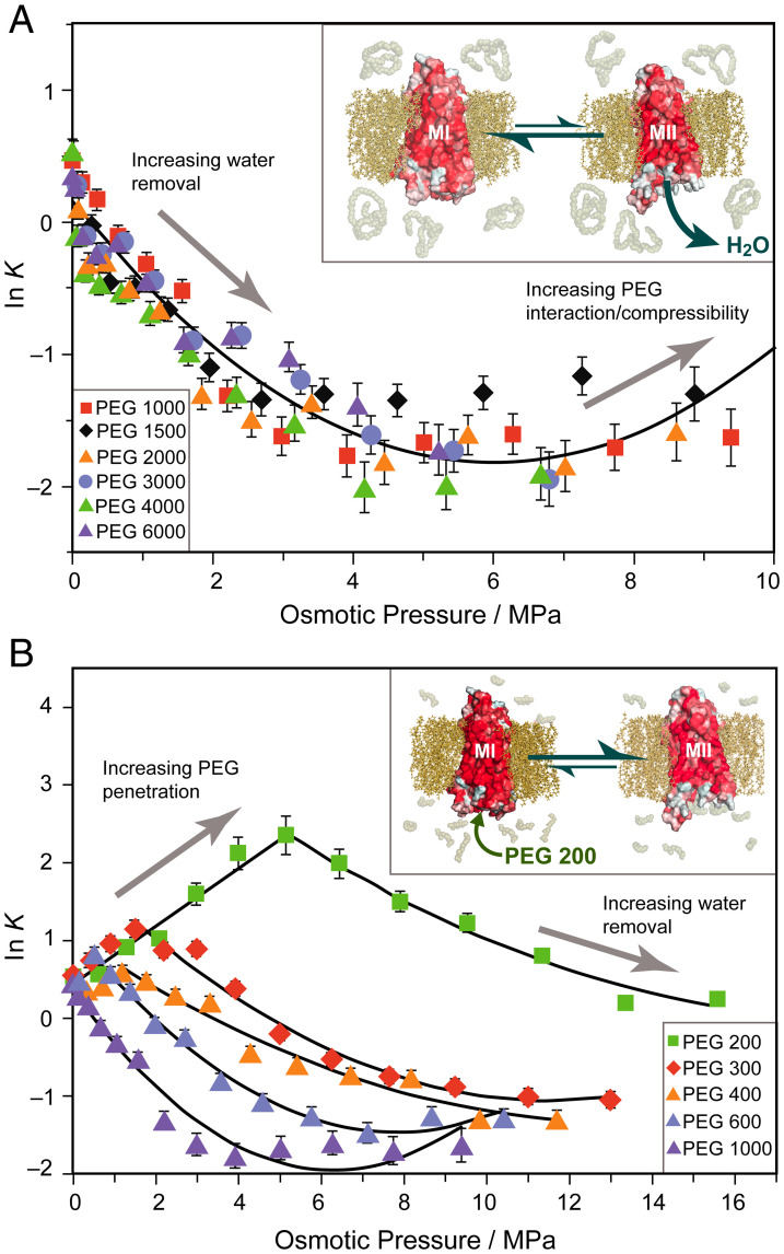
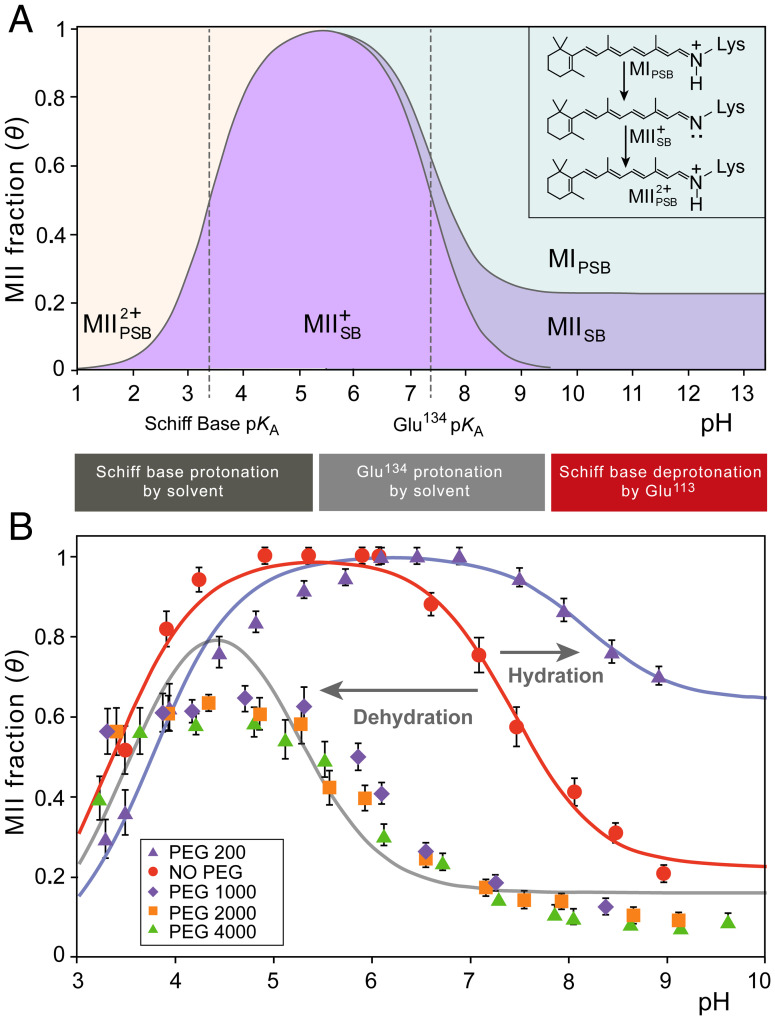
The Rhodopsin family of G-protein–coupled receptors (GPCRs) comprises the targets of nearly a third of all pharmaceuticals. Despite structural water present in GPCR X-ray structures, the physiological relevance of these solvent molecules to rhodopsin signaling remains unknown. Here, we show experimental results consistent with the idea that rhodopsin activation in lipid membranes is coupled to bulk water movements into the protein. To quantify hydration changes, we measured reversible shifting of the metarhodopsin equilibrium due to osmotic stress using an extensive series of polyethylene glycol (PEG) osmolytes. We discovered clear evidence that light activation entails a large influx of bulk water (∼80–100 molecules) into the protein, giving insight into GPCR activation mechanisms. Various size polymer osmolytes directly control rhodopsin activation, in which large solutes are excluded from rhodopsin and dehydrate the protein, favoring the inactive state. In contrast, small osmolytes initially forward shift the activation equilibrium until a quantifiable saturation point is reached, similar to gain-of-function protein mutations. For the limit of increasing osmolyte size, a universal response of rhodopsin to osmotic stress is observed, suggesting it adopts a dynamic, hydrated sponge-like state upon photoactivation. Our results demand a rethinking of the role of water dynamics in modulating various intermediates in the GPCR energy landscape. We propose that besides bound water, an influx of bulk water plays a necessary role in establishing the active GPCR conformation that mediates signaling.

Keywords: GPCR; osmotic stress; rhodopsin; sponge model; structural water.


Conflict of interest statement

The authors declare no competing interest.

Figures

Fig. 1.
Fig. 1.
Rhodopsin activation in lipid membranes entails a large influx of bulk water into the protein core. (A) Dark-state rhodopsin has a closed, dehydrated conformation of the 7-transmembrane helical bundle (30). (B) Open metarhodopsin-II conformation is adopted after retinal photoisomerization with outward-tilted TM6 (blue) and extended TM5 (green) helices following pH-dependent breakage of the Glu134–Arg135 salt bridge (31). (C) Large numbers of water molecules (~80–100) enter rhodopsin as determined by osmotic stress data for various-sized polyethylene glycols (PEGs). Smaller PEGs show smaller apparent water influx than more excluded large PEGs. The number of water molecules determined by the universal large osmolyte response is indicated by the dotted line. (D) Increase in virial coefficient ΔC for volume versus osmotic pressure accompanies rhodopsin photoactivation. Larger ΔC is determined in the exclusion limit of larger PEGs with the dotted line as the universal response. (E) Viewed from the intracellular side, dark-state rhodopsin is closed with residues colored by hydrophobicity (red, hydrophobic; white, hydrophilic). (F) The MII state shows an opened hydrophilic cavity (white) to accommodate the influx of bulk water which (G) widens as the C-terminal peptide of the G-protein (here Gi) is docked (8).
Fig. 2.
Fig. 2.
UV-visible difference spectroscopy reveals shift in equilibrium to inactive MI state upon osmotic dehydration. (A) Experimental difference UV-visible spectra of rhodopsin (light-activated minus dark state) show mixtures of inactive MI and active MII at varying pH conditions. (B) Singular-value decomposition yields two major singular vectors corresponding to experimental MI and MII basis spectra isolated by temperature and pH (pH 9.2 and 5 °C for MI, pH 5.0 and 15 °C for MII). (C) Increasing concentrations of osmolyte PEG 1000 shift spectrum toward MI component. (D) For large-PEG osmolytes the logarithm of the metarhodopsin equilibrium constant (K = [MII]/[MI]) varies approximately quadratically with applied osmotic pressure yielding the change in hydration and virial coefficient C for rhodopsin activation under standard-state conditions.
Fig. 3.
Fig. 3.
Large and small molar mass osmolytes affect rhodopsin differently and show emergence of a universal trend for excluded polymers. (A) Natural logarithm of the MI–MII equilibrium constant (K = [MII]/[MI]) has approximately second-order relationship to osmotic pressure of large PEGs. A universal trend arises for PEGs of Mr between 1,000 and 6,000 Da with the linear term proportional to change in hydration. Inset: Metarhodopsin equilibrium is shifted to the MI (closed) state by large polymer osmolytes which are entropically excluded and dehydrate the protein. (B) Initially, small osmolytes (PEG 200–PEG 600) forward shift ln K to the MII state. A saturation effect is observed beyond which the equilibrium is back shifted to MI resembling the large osmolyte behavior. As PEG size increases, the trend behaves more like the universal behavior. Inset: Small osmolytes such as PEG 200 penetrate the transducin binding cavity and stabilize the open active MII state until it is saturated with small PEGs.
Fig. 4.
Fig. 4.
Osmotic pressure induces large shifts of pH-dependent activation of rhodopsin. (A) Influences of pH on rhodopsin are described by phenomenological Henderson-Hasselbalch equation involving two pKA values and an alkaline endpoint. The states are distinguished by having a protonated or deprotonated Schiff base (PSB or SB, indicated by a subscript), while a superscript indicates the charge relative to MI. The lower pKA (designated pKA,SB) reflects pH-dependent protonation of the retinal Schiff base (SB), which lowers the apparent MII fraction detected by UV-visible spectroscopy. The higher pKA value (designated pKA,Glu) reflects protonation of Glu134 in the E(D)RY motif to stabilize the fully active MII conformation. The alkaline endpoint at higher pH corresponds to MII substates that persist at higher temperatures even when Glu134 is fully deprotonated. (B) Applied osmotic stress stemming from large osmolytes (50% wt/wt at T = 15 °C) back shifts the rhodopsin activation titration curve from pKA = 7.4 to 5.2. At 30% wt/wt PEG 200 (T = 15 °C) the titration curve is maximally forward shifted to a pKA of 8.2 favoring the active MII state. Also observed is an osmolyte effect on the alkaline endpoint: small osmolytes stabilize the open MII conformation even when Glu134 is fully deprotonated, increasing the alkaline endpoint, whereas dehydrating large osmolytes decrease the deprotonated MII population and thus the alkaline endpoint.

Similar articles

Cited by

References

    1. Stevens R. C., et al. , The GPCR Network: A large-scale collaboration to determine human GPCR structure and function. Nat. Rev. Drug Discov. 12, 25–34 (2013). - PMC - PubMed
    1. Latorraca N. R., Venkatakrishnan A. J., Dror R. O., GPCR dynamics: Structures in motion. Chem. Rev. 117, 139–155 (2017). - PubMed
    1. Hilger D., Masureel M., Kobilka B. K., Structure and dynamics of GPCR signaling complexes. Nat. Struct. Mol. Biol. 25, 4–12 (2018). - PMC - PubMed
    1. Weis W. I., Kobilka B. K., The molecular basis of G protein-coupled receptor activation. Annu. Rev. Biochem. 87, 897–919 (2018). - PMC - PubMed
    1. Venkatakrishnan A. J., et al. , Diverse GPCRs exhibit conserved water networks for stabilization and activation. Proc. Natl. Acad. Sci. U.S.A. 116, 3288–3293 (2019). - PMC - PubMed

Publication types

MeSH terms

LinkOut - more resources