. 2022 May 31;94(21):7619-7627.
doi: 10.1021/acs.analchem.2c00835. Epub 2022 May 18.

Detecting Intact Virus Using Exogenous Oligonucleotide Labels

Affiliations

Detecting Intact Virus Using Exogenous Oligonucleotide Labels

Thomas R Carey et al. Anal Chem. .

Abstract

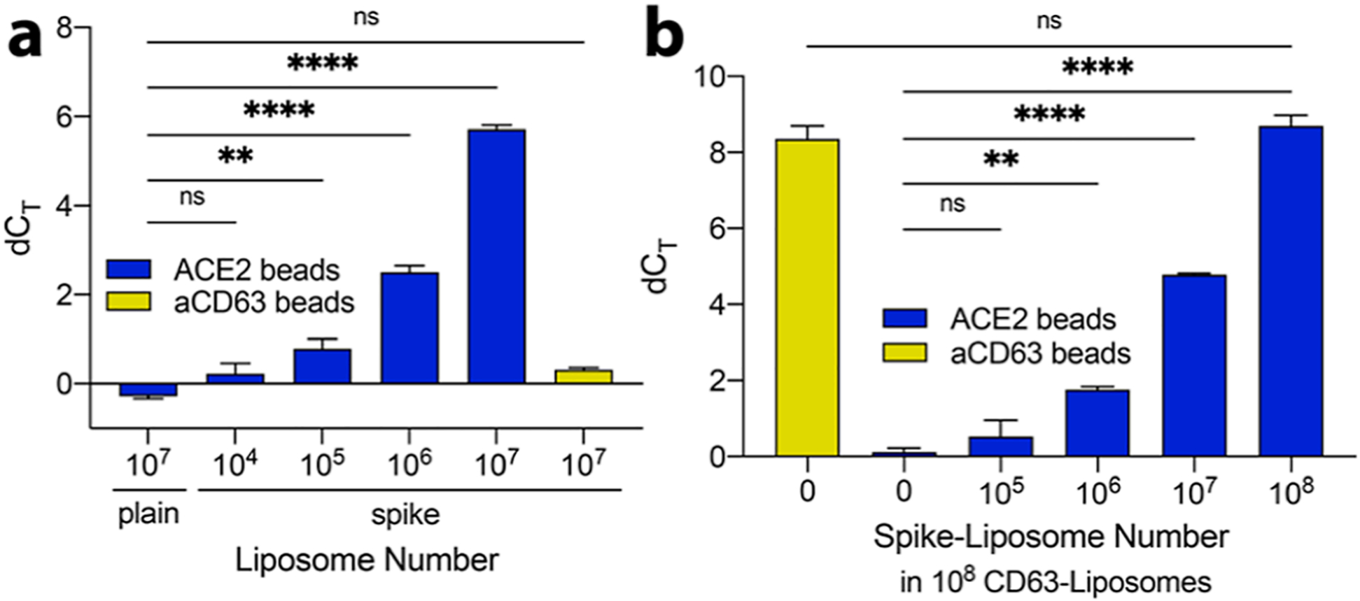
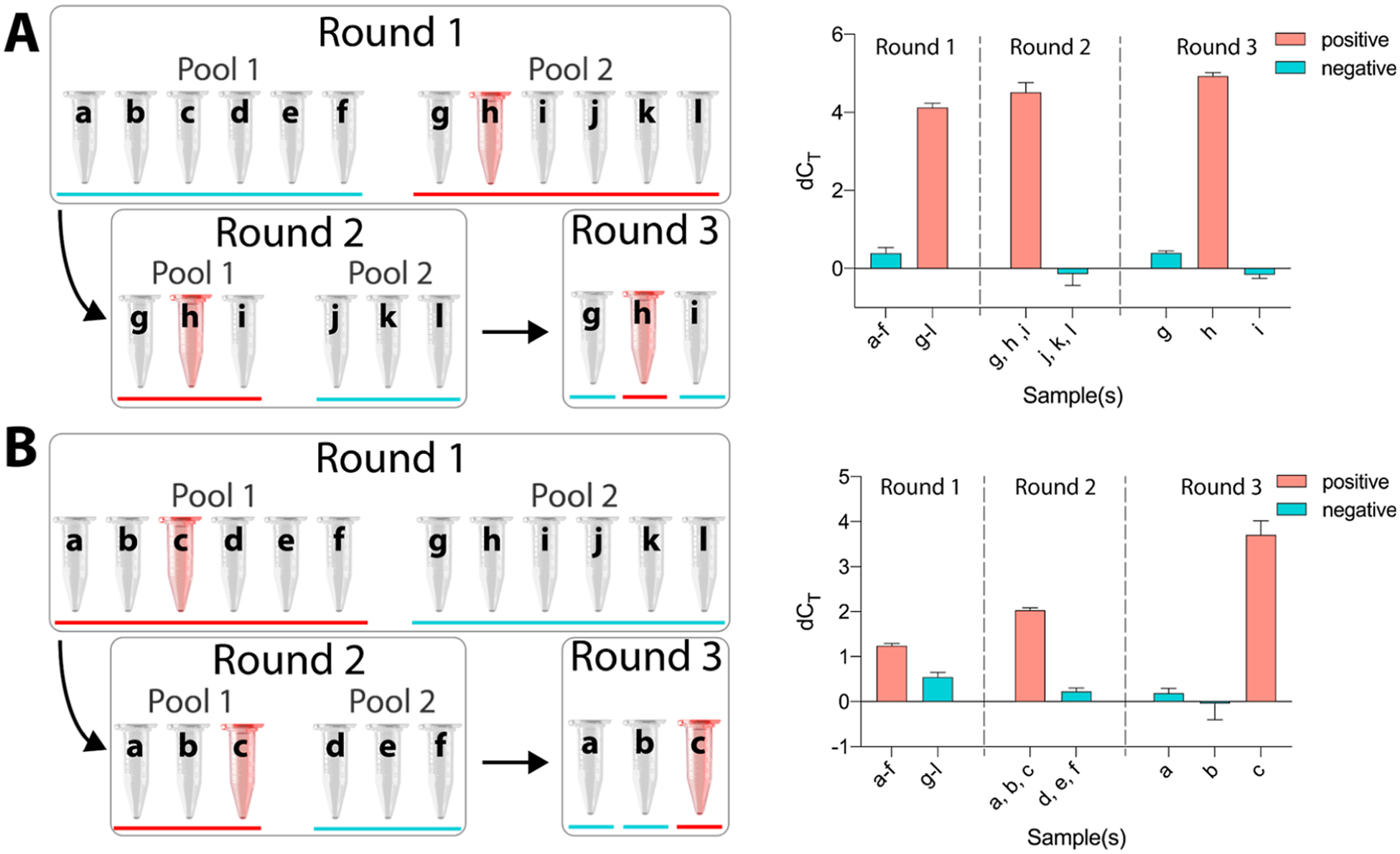
The COVID-19 pandemic has revealed how an emerging pathogen can cause a sudden and dramatic increase in demand for viral testing. Testing pooled samples could meet this demand; however, the sensitivity of reverse transcription quantitative polymerase chain reaction (RT-qPCR), the gold standard, significantly decreases with an increasing number of samples pooled. Here, we introduce detection of intact virus by exogenous-nucleotide reaction (DIVER), a method that quantifies intact virus and is robust to sample dilution. As demonstrated using two models of severe acute respiratory syndrome coronavirus 2, DIVER first tags membraned particles with exogenous oligonucleotides, then captures the tagged particles on beads functionalized with a virus-specific capture agent (in this instance, angiotensin-converting enzyme 2), and finally quantifies the oligonucleotide tags using qPCR. Using spike-presenting liposomes and spike-pseudotyped lentivirus, we show that DIVER can detect 1 × 10⁵ liposomes and 100 plaque-forming units of lentivirus and can successfully identify positive samples in pooling experiments. Overall, DIVER is well positioned for efficient sample pooling and clinical validation.


Conflict of interest statement

The authors declare the following competing financial interest(s): T.R.C., M.K, and L.L.S. have submitted a U.S. patent application (PCT/US20/62957) involving the method to detect via qPCR lipid bilayer nanoparticles labeled with cholesterol-modified oligonucleotides.

Figures

Figure 1.
Figure 1.
(a) Workflow for isolating, labeling, capturing, and quantifying nanoparticles displaying spike protein. Step 1: isolate lipid bilayer nanoparticles <200 nm in diameter using membrane affinity columns; step 2: label isolated nanoparticles, including enveloped viruses and background EVs, with cholesterol-tagged single-stranded DNA (ssDNA) oligonucleotides; step 3: capture ssDNA oligonucleotide-labeled enveloped viruses, or models thereof, on paramagnetic beads functionalized with a capture agent, such as ACE2 for SARS-CoV-2; step 4: wash to remove unbound oligonucleotides and background EVs; step 5: using qPCR with primers specific to oligonucleotide labels, quantify ssDNA, which is directly correlated to the number of target enveloped virus particles. (b) Details of oligonucleotide labeling scheme in step 2 in (a). A 3′ cholesterol-tagged universal anchor oligonucleotide, prehybridized to a qPCR detection oligonucleotide, self-embeds in the lipid bilayer membrane. A 5′ cholesterol-tagged universal coanchor oligonucleotide subsequently self-embeds into the lipid bilayer membrane and hybridizes with the first 20 nts of the universal anchor, thereby stabilizing the complex.
Figure 2.
Figure 2.
(a) Dilution series of spike liposomes suspended in buffer. Solutions containing ≥105 spike liposomes produced a signal significantly greater than 107 plain liposomes when incubated with ACE2 beads. 107 spike liposomes produced a signal comparable to 107 plain liposomes when incubated with aCD63 beads, indicating that the qPCR signal results specifically from the capture of spike liposomes onto ACE2 beads. (b) Dilution series of spike liposomes in a background of 108 CD63 liposomes. When incubated with ACE2 beads, solutions containing ≥106 spike liposomes produced a signal significantly greater than those containing just 108 CD63 liposomes (zero spike liposomes). As a control, 108 CD63 liposomes with no spike liposomes were incubated with aCD63 beads. The qPCR signal was comparable to that of 108 spike liposomes incubated with ACE2 beads, indicating that the capture of liposomes is highly specific. For all panels, the qPCR signal is presented as dCT, the difference in threshold cycle between each sample and a no-liposome control; error bars represent SEM; ns: not significant; *: p < 0.05, **: p < 0.01, ****: p < 0.0001, one-way ANOVA with Dunnett’s (a) or Bonferroni’s (b) multiple comparison test.
Figure 3.
Figure 3.
(a) qPCR signal, where dCT is the difference in threshold cycle between lentivirus samples and a no-virus control, was significantly greater for 100 pfu spike-pseudotyped lentivirus than 100 pfu VSV-G-pseudotyped lentivirus captured onto ACE2 beads. Conversely, the qPCR signal was significantly greater for 100 pfu VSV-G-pseudotyped lentivirus than that for 100 pfu spike-pseudotyped lentivirus captured onto aVSV-G beads. aCD63 beads were employed to capture background EVs. Signals from spike- and VSV-G-pseudotyped samples did not significantly differ from one another or from those of no-virus control samples, confirming that similar numbers of EVs were present in spike- and VSV-G-pseudotyped lentivirus and no-virus control cell culture supernatants. **: p < 0.01, *: p < 0.05, nested t-test to compare spike-pseudotyped and VSV-G-pseudotyped lentivirus for each marker, n = 4, each with n = 3 technical replicates. (b) RT-qPCR quantification of lentivirus indicates that 100 pfu spike-pseudotyped lentivirus corresponds to an RNA copy number of approximately 9816, while 100 pfu VSV-G-pseudotyped lentivirus corresponds to an RNA copy number of approximately 8365; n = 2. For all panels, error bars represent SEM.
Figure 4.
Figure 4.
Sample pooling enables a substantial reduction in resource usage for COVID-19 testing. In the pooling scheme shown here, the total number of qPCR tests is reduced from 12 to 7, a 42% reduction. A red bar indicates a pool or sample that tests positive, and a red tube indicates a positive sample. (A) Blinded demonstration of sample pooling using DIVER with liposomes. Twelve samples were prepared, each containing 107 CD63 liposomes/μL; one sample also contained 106 spike liposomes/μL. In round 1, 10 μL of each sample was combined into pools of six. In round 2, for the pool that was identified as positive, 10 μL of each sample was combined into pools of three. Finally, in round 3, 10 μL of each sample in the positive pool was analyzed individually. DIVER correctly identified sample h as containing 106 spike liposomes/μL. The qPCR signal is presented here as dCT, the difference in threshold cycle between each pool or sample and an equal-volume no-liposome control. (B) Blinded demonstration of sample pooling using DIVER with spike-pseudotyped lentivirus. Similar to the pooling demonstration with liposomes in (A), 12 samples were prepared, each of which contained 4.02 × 10⁶/μL cell-culture-derived EVs, and one sample also contained 20 pfu/μL spike-pseudotyped lentivirus. The same procedure described in (A) was used to correctly identify sample c as containing 20 pfu/μL lentivirus. Here, dCT is the difference in threshold cycle between each pool or sample and a no-virus control. Error bars represent SEM. All other samples were separately confirmed to be negative (Figure S7a,b).

Update of

Similar articles

References

    1. Ritchie H; Mathieu E; Rodés-Guirao L; Appel C; Giattino C; Ortiz-Ospina E; Hasell J; Macdonald B; Beltekian D; Roser M Coronavirus Pandemic (COVID-19). https://ourworldindata.org/covid-vaccinations (accessed Sept 01, 2021).
    1. Cleary B; Hay JA; Blumenstiel B; Harden M; Cipicchio M; Bezney J; Simonton B; Hong D; Senghore M; Sesay AK; Gabriel S; Regev A; Mina MJ Sci. Transl. Med 2021, 13, No. eabf1568. - PMC - PubMed
    1. Schulte PA; Weissman DN; Luckhaupt SE; de Perio MA; Beezhold D; Piacentino JD; Radonovich LJ; Hearl FJ; Howard J J. Occup. Environ. Med 2021, 63, 1–9. - PMC - PubMed
    1. Grobe N; Cherif A; Wang X; Dong Z; Kotanko P Clin. Microbiol. Infect 2021, 27, 1212–1220. - PMC - PubMed
    1. Deka S; Kalita DJ Lab. Physicians 2020, 12, 212–218. - PMC - PubMed

Publication types