. 2022 May 17;39(7):110811.
doi: 10.1016/j.celrep.2022.110811.

The ciliary gene INPP5E confers dorsal telencephalic identity to human cortical organoids by negatively regulating Sonic hedgehog signaling

Affiliations

The ciliary gene INPP5E confers dorsal telencephalic identity to human cortical organoids by negatively regulating Sonic hedgehog signaling

Leah Schembs et al. Cell Rep. .

Abstract

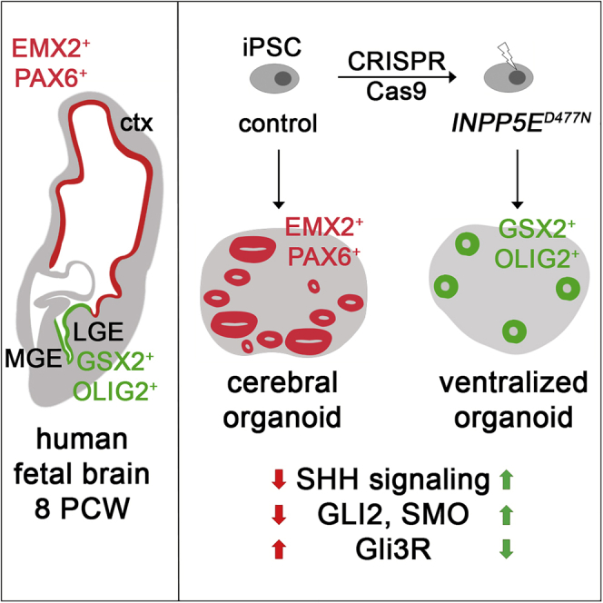
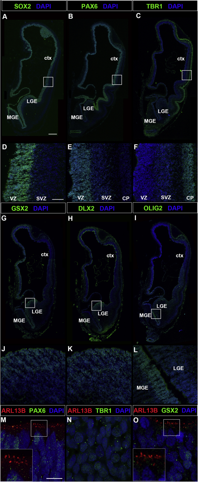
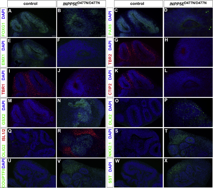
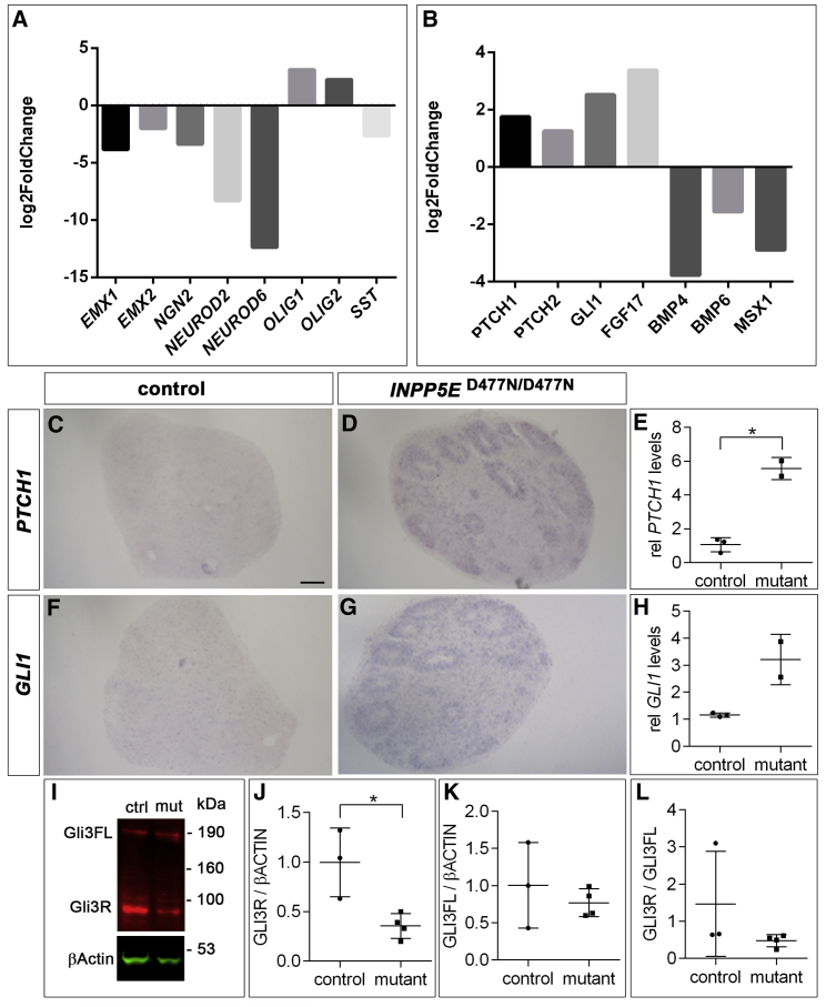
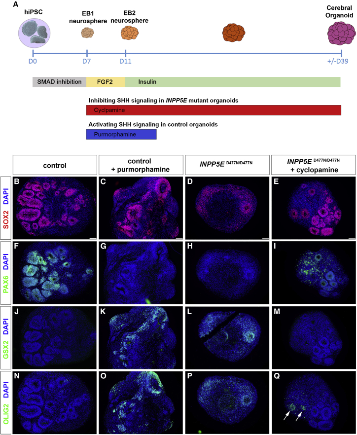
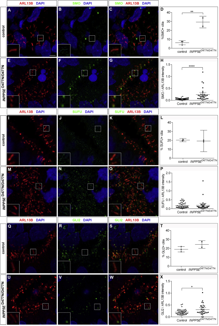
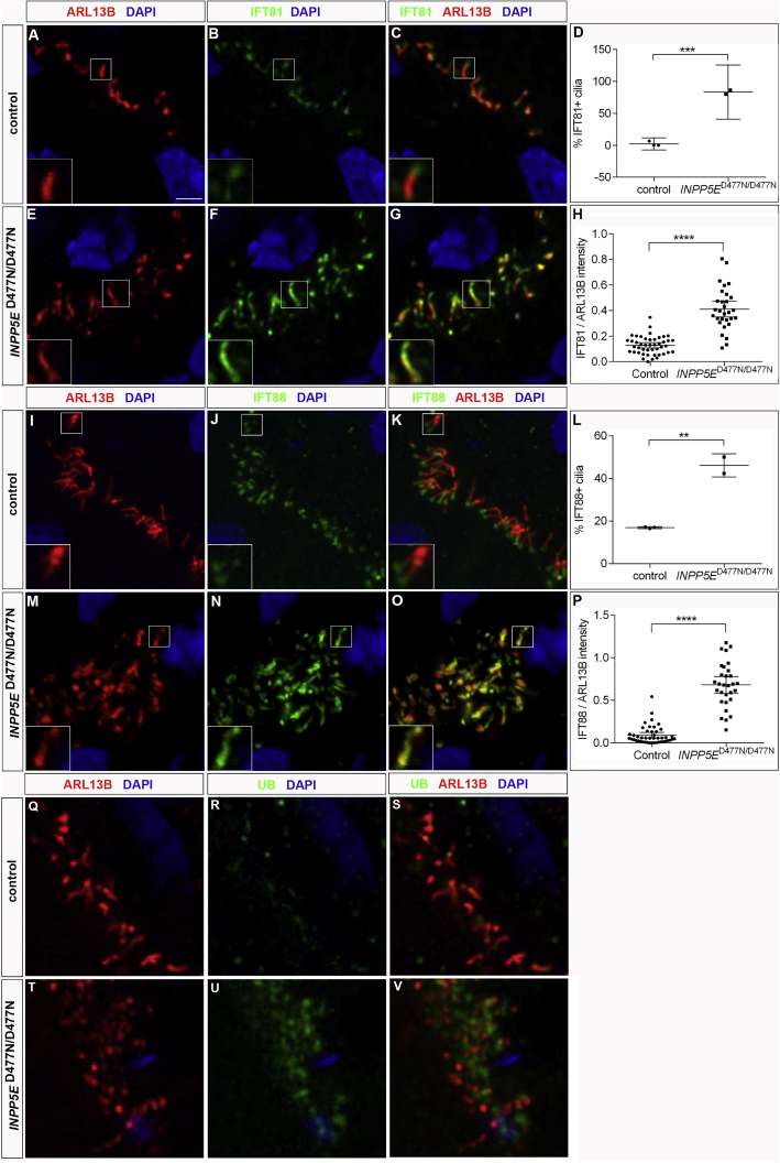
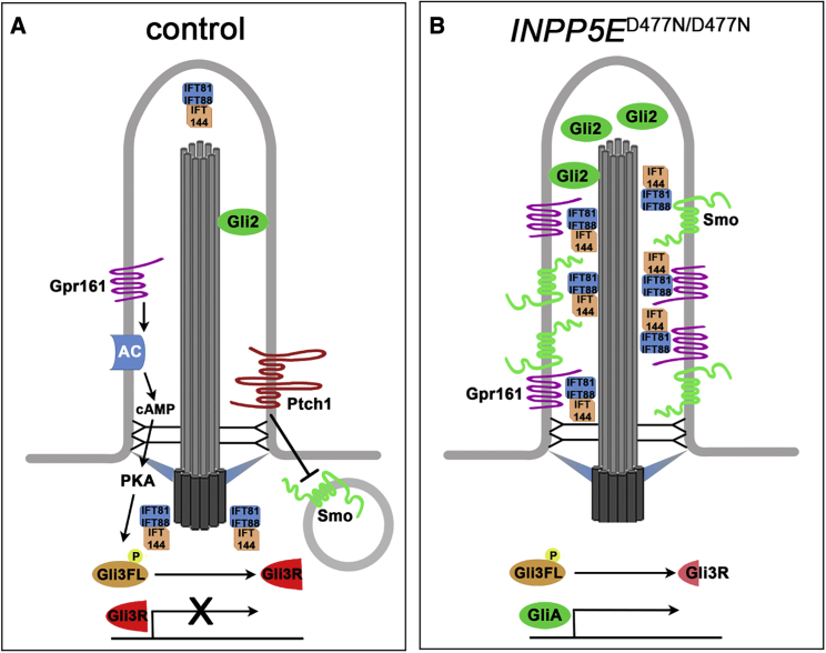
Defects in primary cilia, cellular antennas that control multiple intracellular signaling pathways, underlie several neurodevelopmental disorders, but it remains unknown how cilia control essential steps in human brain formation. Here, we show that cilia are present on the apical surface of radial glial cells in human fetal forebrain. Interfering with cilia signaling in human organoids by mutating the INPP5E gene leads to the formation of ventral telencephalic cell types instead of cortical progenitors and neurons. INPP5E mutant organoids also show increased Sonic hedgehog (SHH) signaling, and cyclopamine treatment partially rescues this ventralization. In addition, ciliary expression of SMO, GLI2, GPR161, and several intraflagellar transport (IFT) proteins is increased. Overall, these findings establish the importance of primary cilia for dorsal and ventral patterning in human corticogenesis, indicate a tissue-specific role of INPP5E as a negative regulator of SHH signaling, and have implications for the emerging roles of cilia in the pathogenesis of neurodevelopmental disorders.

Keywords: CP: Neuroscience; INPP5E; dorsal and ventral patterning; human cortex; primary cilia; sonic hedgehog; telencephalon.


Conflict of interest statement

Declaration of interests The authors declare no competing interests.

Figures

None
Graphical abstract
Figure 1
Figure 1
Cilia in the developing human telencephalon Coronal sections of the 8 PCW telencephalon immunostained with the indicated markers. (A and D) SOX2 expression in dorsal and ventral telencephalic progenitors. (B, C, E, and F) PAX6 and TBR1 expression are confined to dorsal progenitors and neurons, respectively. (G–L) GSX2 (G and J), DLX2 (H and K), and OLIG2 (I and L) expression are confined to the ventral telencephalon. (M–O) ARL13B+ cilia projecting from progenitor cells into the ventricular lumen in the dorsal (M) and ventral (O) telencephalon. Note the absence of ARL13B expression in TBR1+ projection neurons (N). ctx, cortex; LGE, lateral ganglionic eminence; MGE, medial ganglionic eminence; SVZ, subventricular zone; VZ, ventricular zone. Scale bars, 500 μm (A), 100 μm (D), and 2.5 μm (M). See also Figure S1.
Figure 2
Figure 2
The INPP5E mutation interferes with telencephalic marker gene expression Control and INPP5ED477N/D477N organoids were immunostained with the indicated markers. (A and B) The telencephalon marker FOXG1 was expressed in organoids of both genotypes. (C–F) The dorsal aRGC markers PAX6 (C and D) and EMX1 (E and F) were expressed in control, but not in mutant, organoids. (G and H) TBR2-labeled basal progenitor cells that were largely absent in INPP5ED477N/D477N organoids. (I–L) Expression of the cortical neuron markers TBR1 and CTIP2 was largely absent in mutant organoids. (M–R) Expression of the ventral telencephalic progenitor markers GSX2 (M and N), DLX2 (O and P), and OLIG2 (Q and R) in mutant, but not in control, organoids. ISL1/2 labeling spiny medium interneurons was detected in INPP5E mutant organoids (Q and R). (S–X) Expression analyses of the ventral telencephalic interneuron markers NKX2.1 (S and T), COUPTFII (U and V), and SST (W and X). Scale bars, 100 μm (A) and 50 μm (B). See also Figure S3.
Figure 3
Figure 3
SHH signaling is up-regulated in INPP5ED477N/D477N organoids (A and B) Examples of differentially expressed dorsal and ventral (A) and signaling factor genes (B) with indicated log2Fold changes in D24 organoids. (C, D, F, and G) In situ hybridization to detect PTCH1 and GLI1 expression in D39 control and INPP5E mutant organoids. (E and H) Representative example of qRT-PCR analyses showing PTCH1 (E) and GLI1 (H) mRNA expression relative to ATP5 at D24. (I) GLI3 western blot on D24 organoid tissue revealed the GLI3 full-length (FL) and repressor (R) forms. (J–L) Quantification of GLI3 western blot. GLI3R levels are decreased in INPP5ED477N/D477N organoids, while GLI3FL levels and the GLI3R/GLI3FL ratio (L) are not affected. All statistical data are presented as means ± SD; unpaired t tests with Welch’s correction (n = 3 for control and n = 2 for mutant; E and H); unpaired t tests (J and K); Mann-Whitney test (L) with n = 3 for control and n = 4 for mutant; p < 0.05. Scale bar, 100 μm (C). See also Figures S4 and S5.
Figure 4
Figure 4
SHH signaling is necessary and sufficient to ventralize cortical organoids (A) Experimental protocol to either block or ectopically activate SHH signaling in cortical organoids. (B–Q) Organoids stained with the indicated antibodies after treatment with purmorphamine (C, G, K, and O) and cyclopamine (E, I, M, and Q). (B–E) SOX2 expression revealed neuroepithelia. (F–I) PAX6 expression is reduced in purmorphamine-treated control organoids but up-regulated after inhibiting SHH signaling in INPP5E mutant organoids. (J–Q) GSX2 and OLIG2 expression occurs in only a few cells in control organoids (J and N) but is up-regulated after activating SHH signaling (K and O) or in untreated mutant organoids (L and P). (M and Q) Cyclopamine treatment inhibits GSX2 and OLIG2 expression in mutant organoids except for a few cells (arrows in Q). Scale bars, 100 μm (B–E).
Figure 5
Figure 5
Ciliary expression of SHH signaling components Control and INPP5ED477N/D477N organoids were immunostained with the indicated markers. (A–H) SMO was expressed in a higher proportion of cilia and at higher levels in INPP5ED477N/D477N organoids. (I–P) There were no significant changes either in the proportion of positive cilia or in the expression levels for SUFU. (Q–X) GLI2 accumulated in mutant cilia. Statistical data are presented as means ± 95% confidence intervals (CIs); unpaired t tests (D, L, and T) and Mann-Whitney tests (H, P, and X); n = 3 (control) and n = 2 (mutant) lines for (D), (L), and (T); n = 45 (control) and n = 30 (mutant) cilia from three and two different lines, respectively (H, P, and X); p < 0.05; ∗∗p < 0.01; ∗∗∗∗p < 0.0001. Scale bar, 2.5 μm. See also Figures S6 and S7.
Figure 6
Figure 6
IFT-B proteins in INPP5ED477N/D477N organoids Control and INPP5ED477N/D477N organoids were immunostained with the indicated markers. (A–P) IFT81 and IFT88 were expressed in a higher proportion of ciliary axonemes and at increased levels. (Q–V) UB expression increased at the base, but not in the axoneme, of INPP5ED477N/D477N cilia. Statistical data are presented as means ± 95% CIs; unpaired t tests (D and L); unpaired t test with Welch’s correction (H) and Mann-Whitney tests (P); n = 3 (control) and n = 2 (mutant) lines for (D), (L), and (T); n = 45 (control) and n = 30 (mutant) cilia from three and two different lines, respectively (H, P, and X); ∗∗p < 0.01; ∗∗∗p < 0.001; ∗∗∗∗p < 0.0001. Scale bar, 2.5 μm. See also Figures S6 and S7.
Figure 7
Figure 7
Schematic summary of the changes in the activity and localization of SHH signaling components identified in INPP5E mutant cortical organoids (A) In control organoids, SMO ciliary levels are low and the GLI3 repressor form (GLI3R) suppressing SHH signaling predominates. (B) In INPP5E mutant organoids, the ciliary localization of several negative and positive regulators of SHH signaling is disturbed. SMO, GLI2, GPR161, and several IFT proteins accumulate in the cilium. GLI3R levels are reduced, and SHH target genes are activated.

Similar articles

Cited by

References

    1. Abbate J., Lacayo J.C., Prichard M., Pari G., McVoy M.A. Bifunctional protein conferring enhanced green fluorescence and puromycin resistance. Biotechniques. 2001;31:336–340. doi: 10.2144/01312st05. - DOI - PubMed
    1. Andreu-Cervera A., Catala M., Schneider-Maunoury S. Cilia, ciliopathies and hedgehog-related forebrain developmental disorders. Neurobiol. Dis. 2021;150:105236. doi: 10.1016/j.nbd.2020.105236. - DOI - PubMed
    1. Arlotta P., Pasca S.P. Cell diversity in the human cerebral cortex: from the embryo to brain organoids. Curr. Opin. Neurobiol. 2019;56:194–198. doi: 10.1016/j.conb.2019.03.001. - DOI - PubMed
    1. Ashique A.M., Choe Y., Karlen M., May S.R., Phamluong K., Solloway M.J., Ericson J., Peterson A.S. The Rfx4 transcription factor modulates Shh signaling by regional control of ciliogenesis. Sci. Signal. 2009;2:ra70. doi: 10.1126/scisignal.2000602. - DOI - PubMed
    1. Bagley J.A., Reumann D., Bian S., Levi-Strauss J., Knoblich J.A. Fused cerebral organoids model interactions between brain regions. Nat. Methods. 2017;14:743–751. doi: 10.1038/nmeth.4304. - DOI - PMC - PubMed

Publication types

Substances