Review
. 2022 Aug 22:42:91-113.
doi: 10.1146/annurev-nutr-062220-105200. Epub 2022 May 18.

Reprogramming of Hepatic Metabolism and Microenvironment in Nonalcoholic Steatohepatitis

Affiliations
Review

Reprogramming of Hepatic Metabolism and Microenvironment in Nonalcoholic Steatohepatitis

Liangyou Rui et al. Annu Rev Nutr. .

Abstract

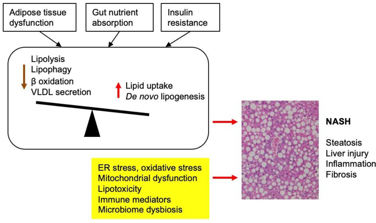
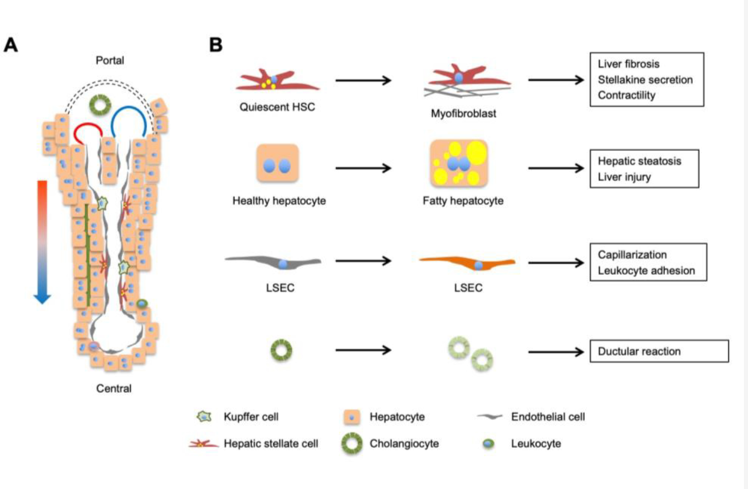
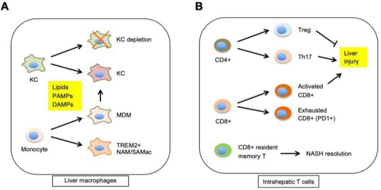
Nonalcoholic fatty liver disease (NAFLD), a spectrum of metabolic liver disease associated with obesity, ranges from relatively benign hepatic steatosis to nonalcoholic steatohepatitis (NASH). The latter is characterized by persistent liver injury, inflammation, and liver fibrosis, which collectively increase the risk for end-stage liver diseases such as cirrhosis and hepatocellular carcinoma. Recent work has shed new light on the pathophysiology of NAFLD/NASH, particularly the role of genetic, epigenetic, and dietary factors and metabolic dysfunctions in other tissues in driving excess hepatic fat accumulation and liver injury. In parallel, single-cell RNA sequencing studies have revealed unprecedented details of the molecular nature of liver cell heterogeneity, intrahepatic cross talk, and disease-associated reprogramming of the liver immune and stromal vascular microenvironment. This review covers the recent advances in these areas, the emerging concepts of NASH pathogenesis, and potential new therapeutic opportunities.

Keywords: NAFLD; NASH; fatty liver; lipid metabolism; microenvironment; single cell; steatosis.


Conflict of interest statement

The authors declare no conflict of financial interest.

Figures

Figure 1.
Figure 1.. An overview of the pathophysiological mechanisms underlying NASH.
Hepatic steatosis results from imbalance between lipid supply (dietary lipids, adipose lipolysis and de novo lipogenesis) and utilization (fatty acid β oxidation and VLDL secretion) by hepatocytes. Insulin resistance, adipose tissue dysfunction, and excess dietary fructose drive hepatic steatosis, which predisposes hepatocytes to injury and cell death in the presence of other stress signals and pathogenic insults. Liver injury triggers remodeling of the immune and stromal vascular microenvironment that results in chronic tissue inflammation and liver fibrosis.
Figure 2.
Figure 2.. Regulation of hepatic de novo lipogenesis.
Hepatic DNL gene program is regulated by nutrients, hormonal cues, and pathophysiological signals, which act on several transcription factors and chromatin regulators that culminate in lipogenic gene induction. Carbohydrates such as fructose stimulate gene expression and transcriptional activity of ChREBP, whereas insulin and LXR activation leads to SREBP1c-mediated lipogenic induction. ER stress triggers activation of XBP1 and SREBP1c to regulate lipogenic gene expression.
Figure 3.
Figure 3.. Liver cell heterogeneity and reprogramming during NASH.
(A) A liver lobule contains parenchymal hepatocytes (60–70% of liver cells) and non-parenchymal cells (30–40%), including sinusoidal endothelial cells, hepatic stellate cells, cholangiocytes, and immune cells (Kupffer cells and leukocytes). Mixed blood from hepatic artery (red) and portal vein (blue) flows along the portal to central direction, creating gradients of oxygen, nutrients, hormones, and gut-derived factors that drive transcriptomic and functional zonation of cells in the liver. (B) Reprogramming of major cell types during NASH pathogenesis. Individual cell types in the liver undergo cell type-specific transcriptomic, metabolic, and functional reprogramming during NASH progression.
Figure 4.
Figure 4.. Reprogramming of macrophages and intrahepatic T cells during NASH.
(A) NASH triggers infiltration of circulating monocytes into the liver, a subset of which polarize to a distinct macrophage subtype characterized by high levels of Trem2 expression (NASH-associated macrophage, NAM or scar-associated macrophage, SAMac). Monocyte-derived macrophages (MDM) contribute to the replenishment of depleted Kupffer cells during NASH. The transcriptomic and functional reprogramming of macrophages is regulated by exposure to lipids, PAMPs, and DAMPs. (B) Features of intrahepatic T cell reprogramming during NASH. Different T cell subtypes contribute to liver injury during NASH, fibrosis resolution, and response to immunotherapy agents.

References

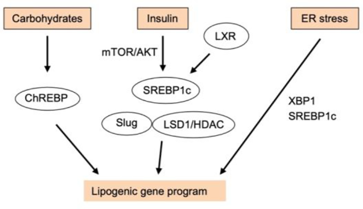
    1. Abdul-Wahed A, Guilmeau S, Postic C. 2017. Sweet Sixteenth for ChREBP: Established Roles and Future Goals. Cell Metab 26: 324–41 - PubMed
    1. Aizarani N, Saviano A, Sagar Mailly L, Durand S, et al. 2019. A human liver cell atlas reveals heterogeneity and epithelial progenitors. Nature 572: 199–204 - PMC - PubMed
    1. Andres-Hernando A, Orlicky DJ, Kuwabara M, Ishimoto T, Nakagawa T, et al. 2020. Deletion of Fructokinase in the Liver or in the Intestine Reveals Differential Effects on Sugar-Induced Metabolic Dysfunction. Cell Metab 32: 117–27 e3 - PMC - PubMed
    1. Arendt BM, Comelli EM, Ma DW, Lou W, Teterina A, et al. 2015. Altered hepatic gene expression in nonalcoholic fatty liver disease is associated with lower hepatic n-3 and n-6 polyunsaturated fatty acids. Hepatology 61: 1565–78 - PubMed
    1. Arriazu E, Ge X, Leung TM, Magdaleno F, Lopategi A, et al. 2017. Signalling via the osteopontin and high mobility group box-1 axis drives the fibrogenic response to liver injury. Gut 66: 1123–37 - PMC - PubMed

Publication types

MeSH terms